Calcium »
PDB 1fn7-1fzc »
1fza »
Calcium in PDB 1fza: Crystal Structure of Fibrinogen Fragment D
Protein crystallography data
The structure of Crystal Structure of Fibrinogen Fragment D, PDB code: 1fza
was solved by
G.Spraggon,
S.J.Everse,
R.F.Doolittle,
with X-Ray Crystallography technique. A brief refinement statistics is given in the table below:
| Resolution Low / High (Å) | 30.00 / 2.90 |
| Space group | P 1 21 1 |
| Cell size a, b, c (Å), α, β, γ (°) | 107.720, 48.080, 167.560, 90.00, 105.70, 90.00 |
| R / Rfree (%) | 26.3 / 36.3 |
Calcium Binding Sites:
The binding sites of Calcium atom in the Crystal Structure of Fibrinogen Fragment D
(pdb code 1fza). This binding sites where shown within
5.0 Angstroms radius around Calcium atom.
In total 2 binding sites of Calcium where determined in the Crystal Structure of Fibrinogen Fragment D, PDB code: 1fza:
Jump to Calcium binding site number: 1; 2;
In total 2 binding sites of Calcium where determined in the Crystal Structure of Fibrinogen Fragment D, PDB code: 1fza:
Jump to Calcium binding site number: 1; 2;
Calcium binding site 1 out of 2 in 1fza
Go back to
Calcium binding site 1 out
of 2 in the Crystal Structure of Fibrinogen Fragment D
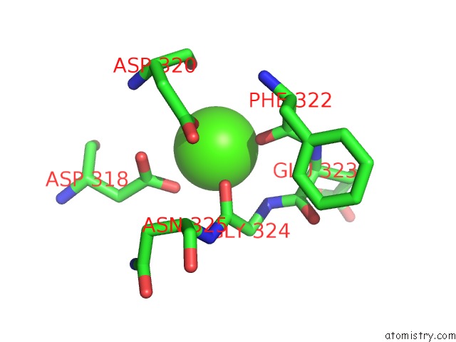
Mono view
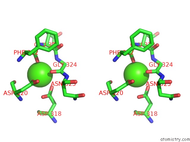
Stereo pair view
Mono view
Stereo pair view
A full contact list of Calcium with other atoms in the Ca binding
site number 1 of Crystal Structure of Fibrinogen Fragment D within 5.0Å range:
|
Calcium binding site 2 out of 2 in 1fza
Go back to
Calcium binding site 2 out
of 2 in the Crystal Structure of Fibrinogen Fragment D
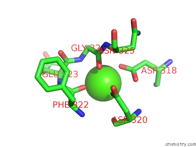
Mono view
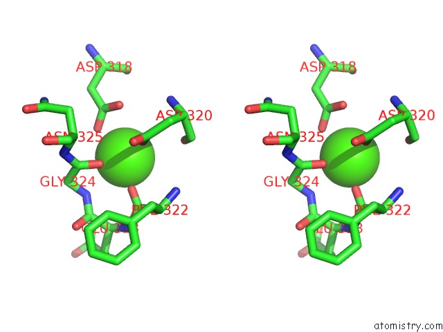
Stereo pair view
Mono view
Stereo pair view
A full contact list of Calcium with other atoms in the Ca binding
site number 2 of Crystal Structure of Fibrinogen Fragment D within 5.0Å range:
|
Reference:
G.Spraggon,
S.J.Everse,
R.F.Doolittle.
Crystal Structures of Fragment D From Human Fibrinogen and Its Crosslinked Counterpart From Fibrin. Nature V. 389 455 1997.
ISSN: ISSN 0028-0836
PubMed: 9333233
DOI: 10.1038/38947
Page generated: Thu Jul 11 08:21:50 2024
ISSN: ISSN 0028-0836
PubMed: 9333233
DOI: 10.1038/38947
Last articles
Zn in 9MJ5Zn in 9HNW
Zn in 9G0L
Zn in 9FNE
Zn in 9DZN
Zn in 9E0I
Zn in 9D32
Zn in 9DAK
Zn in 8ZXC
Zn in 8ZUF