Calcium »
PDB 1fzd-1g8f »
1g1r »
Calcium in PDB 1g1r: Crystal Structure of P-Selectin Lectin/Egf Domains Complexed with Slex
Protein crystallography data
The structure of Crystal Structure of P-Selectin Lectin/Egf Domains Complexed with Slex, PDB code: 1g1r
was solved by
W.S.Somers,
R.T.Camphausen,
with X-Ray Crystallography technique. A brief refinement statistics is given in the table below:
| Resolution Low / High (Å) | 14.00 / 3.40 |
| Space group | P 1 21 1 |
| Cell size a, b, c (Å), α, β, γ (°) | 81.140, 60.520, 91.440, 90.00, 103.28, 90.00 |
| R / Rfree (%) | 22.7 / 32.2 |
Calcium Binding Sites:
The binding sites of Calcium atom in the Crystal Structure of P-Selectin Lectin/Egf Domains Complexed with Slex
(pdb code 1g1r). This binding sites where shown within
5.0 Angstroms radius around Calcium atom.
In total 4 binding sites of Calcium where determined in the Crystal Structure of P-Selectin Lectin/Egf Domains Complexed with Slex, PDB code: 1g1r:
Jump to Calcium binding site number: 1; 2; 3; 4;
In total 4 binding sites of Calcium where determined in the Crystal Structure of P-Selectin Lectin/Egf Domains Complexed with Slex, PDB code: 1g1r:
Jump to Calcium binding site number: 1; 2; 3; 4;
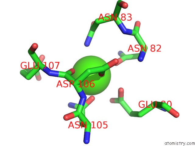
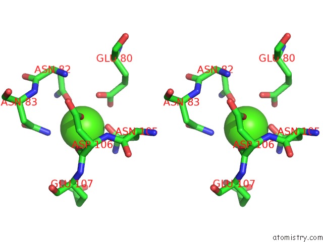
Calcium binding site 1 out of 4 in 1g1r
Go back to
Calcium binding site 1 out
of 4 in the Crystal Structure of P-Selectin Lectin/Egf Domains Complexed with Slex
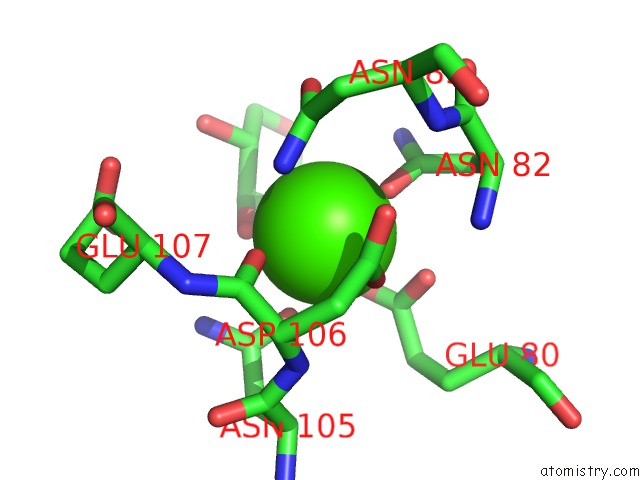
Mono view
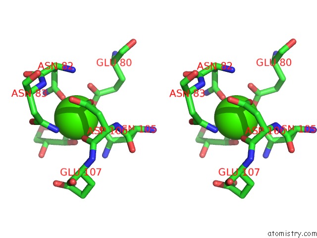
Stereo pair view
Mono view
Stereo pair view
A full contact list of Calcium with other atoms in the Ca binding
site number 1 of Crystal Structure of P-Selectin Lectin/Egf Domains Complexed with Slex within 5.0Å range:
|
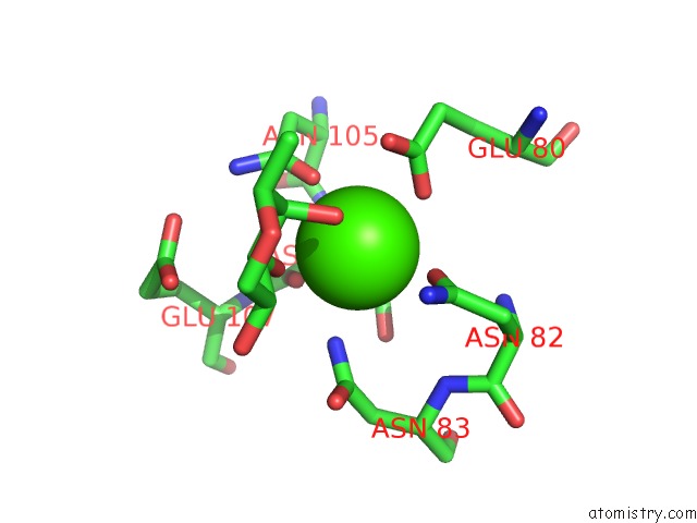
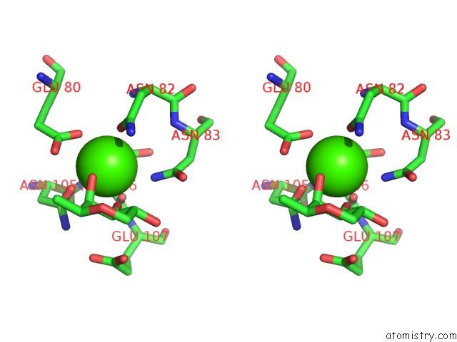
Calcium binding site 2 out of 4 in 1g1r
Go back to
Calcium binding site 2 out
of 4 in the Crystal Structure of P-Selectin Lectin/Egf Domains Complexed with Slex
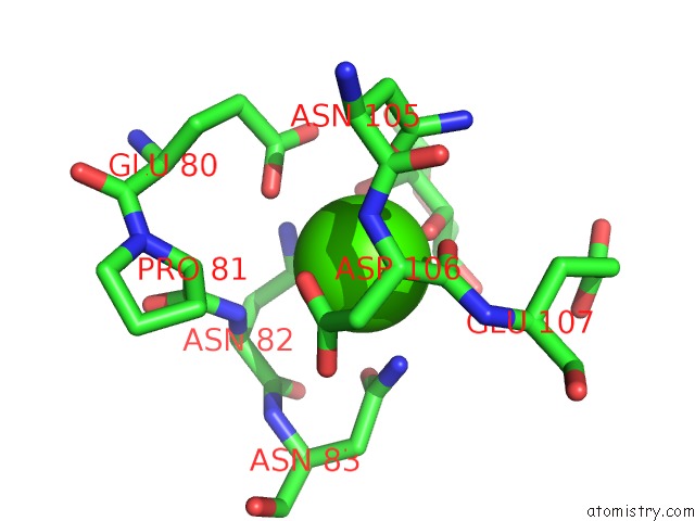
Mono view
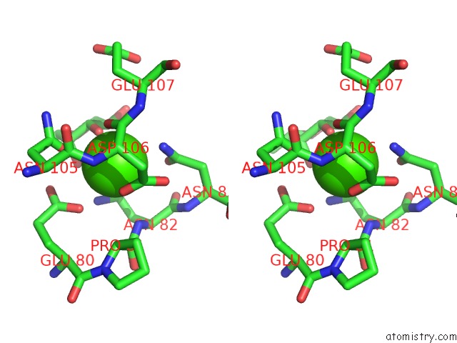
Stereo pair view
Mono view
Stereo pair view
A full contact list of Calcium with other atoms in the Ca binding
site number 2 of Crystal Structure of P-Selectin Lectin/Egf Domains Complexed with Slex within 5.0Å range:
|
Calcium binding site 3 out of 4 in 1g1r
Go back to
Calcium binding site 3 out
of 4 in the Crystal Structure of P-Selectin Lectin/Egf Domains Complexed with Slex
Mono view
Stereo pair view
Mono view
Stereo pair view
A full contact list of Calcium with other atoms in the Ca binding
site number 3 of Crystal Structure of P-Selectin Lectin/Egf Domains Complexed with Slex within 5.0Å range:
|
Calcium binding site 4 out of 4 in 1g1r
Go back to
Calcium binding site 4 out
of 4 in the Crystal Structure of P-Selectin Lectin/Egf Domains Complexed with Slex
Mono view
Stereo pair view
Mono view
Stereo pair view
A full contact list of Calcium with other atoms in the Ca binding
site number 4 of Crystal Structure of P-Selectin Lectin/Egf Domains Complexed with Slex within 5.0Å range:
|
Reference:
W.S.Somers,
J.Tang,
G.D.Shaw,
R.T.Camphausen.
Insights Into the Molecular Basis of Leukocyte Tethering and Rolling Revealed By Structures of P- and E-Selectin Bound to Sle(X) and Psgl-1. Cell(Cambridge,Mass.) V. 103 467 2000.
ISSN: ISSN 0092-8674
PubMed: 11081633
DOI: 10.1016/S0092-8674(00)00138-0
Page generated: Thu Jul 11 08:27:08 2024
ISSN: ISSN 0092-8674
PubMed: 11081633
DOI: 10.1016/S0092-8674(00)00138-0
Last articles
Zn in 9MJ5Zn in 9HNW
Zn in 9G0L
Zn in 9FNE
Zn in 9DZN
Zn in 9E0I
Zn in 9D32
Zn in 9DAK
Zn in 8ZXC
Zn in 8ZUF