Calcium »
PDB 1fzd-1g8f »
1g36 »
Calcium in PDB 1g36: Trypsin Inhibitor Complex
Enzymatic activity of Trypsin Inhibitor Complex
All present enzymatic activity of Trypsin Inhibitor Complex:
3.4.21.4;
3.4.21.4;
Protein crystallography data
The structure of Trypsin Inhibitor Complex, PDB code: 1g36
was solved by
H.Nar,
with X-Ray Crystallography technique. A brief refinement statistics is given in the table below:
| Resolution Low / High (Å) | 8.00 / 1.90 |
| Space group | P 21 21 21 |
| Cell size a, b, c (Å), α, β, γ (°) | 63.100, 69.400, 63.800, 90.00, 90.00, 90.00 |
| R / Rfree (%) | 17.4 / 21.6 |
Calcium Binding Sites:
The binding sites of Calcium atom in the Trypsin Inhibitor Complex
(pdb code 1g36). This binding sites where shown within
5.0 Angstroms radius around Calcium atom.
In total only one binding site of Calcium was determined in the Trypsin Inhibitor Complex, PDB code: 1g36:
In total only one binding site of Calcium was determined in the Trypsin Inhibitor Complex, PDB code: 1g36:
Calcium binding site 1 out of 1 in 1g36
Go back to
Calcium binding site 1 out
of 1 in the Trypsin Inhibitor Complex
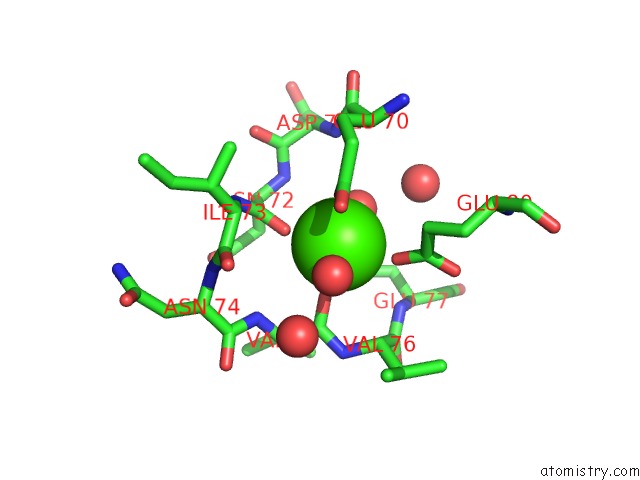
Mono view
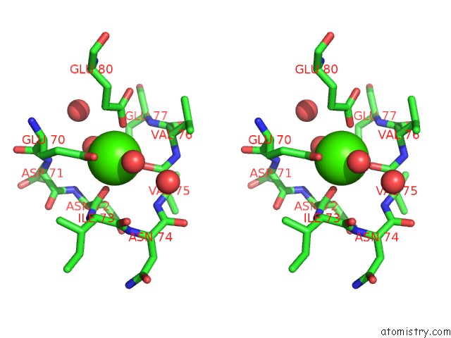
Stereo pair view
Mono view
Stereo pair view
A full contact list of Calcium with other atoms in the Ca binding
site number 1 of Trypsin Inhibitor Complex within 5.0Å range:
|
Reference:
H.Nar,
M.Bauer,
A.Schmid,
J.M.Stassen,
W.Wienen,
H.W.Priepke,
I.K.Kauffmann,
U.J.Ries,
N.H.Hauel.
Structural Basis For Inhibition Promiscuity of Dual Specific Thrombin and Factor Xa Blood Coagulation Inhibitors. Structure V. 9 29 2001.
ISSN: ISSN 0969-2126
PubMed: 11342132
DOI: 10.1016/S0969-2126(00)00551-7
Page generated: Thu Jul 11 08:29:04 2024
ISSN: ISSN 0969-2126
PubMed: 11342132
DOI: 10.1016/S0969-2126(00)00551-7
Last articles
Zn in 9J0NZn in 9J0O
Zn in 9J0P
Zn in 9FJX
Zn in 9EKB
Zn in 9C0F
Zn in 9CAH
Zn in 9CH0
Zn in 9CH3
Zn in 9CH1