Calcium »
PDB 1g8g-1gj6 »
1g9f »
Calcium in PDB 1g9f: Crystal Structure of the Soybean Agglutinin in A Complex with A Biantennary Blood Group Antigen Analog
Protein crystallography data
The structure of Crystal Structure of the Soybean Agglutinin in A Complex with A Biantennary Blood Group Antigen Analog, PDB code: 1g9f
was solved by
L.Buts,
T.W.Hamelryck,
M.-H.Dao-Thi,
R.Loris,
L.Wyns,
M.E.Etzler,
with X-Ray Crystallography technique. A brief refinement statistics is given in the table below:
| Resolution Low / High (Å) | 20.00 / 2.50 |
| Space group | I 41 2 2 |
| Cell size a, b, c (Å), α, β, γ (°) | 122.640, 122.640, 90.560, 90.00, 90.00, 90.00 |
| R / Rfree (%) | 19 / 23 |
Other elements in 1g9f:
The structure of Crystal Structure of the Soybean Agglutinin in A Complex with A Biantennary Blood Group Antigen Analog also contains other interesting chemical elements:
| Manganese | (Mn) | 1 atom |
Calcium Binding Sites:
The binding sites of Calcium atom in the Crystal Structure of the Soybean Agglutinin in A Complex with A Biantennary Blood Group Antigen Analog
(pdb code 1g9f). This binding sites where shown within
5.0 Angstroms radius around Calcium atom.
In total only one binding site of Calcium was determined in the Crystal Structure of the Soybean Agglutinin in A Complex with A Biantennary Blood Group Antigen Analog, PDB code: 1g9f:
In total only one binding site of Calcium was determined in the Crystal Structure of the Soybean Agglutinin in A Complex with A Biantennary Blood Group Antigen Analog, PDB code: 1g9f:
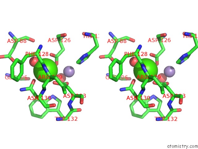
Calcium binding site 1 out of 1 in 1g9f
Go back to
Calcium binding site 1 out
of 1 in the Crystal Structure of the Soybean Agglutinin in A Complex with A Biantennary Blood Group Antigen Analog
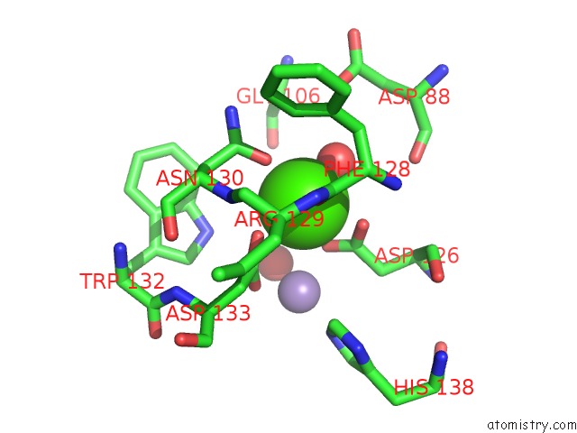
Mono view
Stereo pair view
Mono view
Stereo pair view
A full contact list of Calcium with other atoms in the Ca binding
site number 1 of Crystal Structure of the Soybean Agglutinin in A Complex with A Biantennary Blood Group Antigen Analog within 5.0Å range:
|
Reference:
L.Buts,
M.H.Dao-Thi,
R.Loris,
L.Wyns,
M.Etzler,
T.Hamelryck.
Weak Protein-Protein Interactions in Lectins: the Crystal Structure of A Vegetative Lectin From the Legume Dolichos Biflorus. J.Mol.Biol. V. 309 193 2001.
ISSN: ISSN 0022-2836
PubMed: 11491289
DOI: 10.1006/JMBI.2001.4639
Page generated: Thu Jul 11 08:35:36 2024
ISSN: ISSN 0022-2836
PubMed: 11491289
DOI: 10.1006/JMBI.2001.4639
Last articles
Zn in 9MJ5Zn in 9HNW
Zn in 9G0L
Zn in 9FNE
Zn in 9DZN
Zn in 9E0I
Zn in 9D32
Zn in 9DAK
Zn in 8ZXC
Zn in 8ZUF