Calcium »
PDB 1g8g-1gj6 »
1g9j »
Calcium in PDB 1g9j: X-Tal Structure of the Mutant E44Q of the Cellulase CEL48F in Complex with A Thiooligosaccharide
Enzymatic activity of X-Tal Structure of the Mutant E44Q of the Cellulase CEL48F in Complex with A Thiooligosaccharide
All present enzymatic activity of X-Tal Structure of the Mutant E44Q of the Cellulase CEL48F in Complex with A Thiooligosaccharide:
3.2.1.4;
3.2.1.4;
Protein crystallography data
The structure of X-Tal Structure of the Mutant E44Q of the Cellulase CEL48F in Complex with A Thiooligosaccharide, PDB code: 1g9j
was solved by
G.Parsiegla,
C.Tardif,
J.P.Belaich,
H.Driguez,
R.Haser,
with X-Ray Crystallography technique. A brief refinement statistics is given in the table below:
| Resolution Low / High (Å) | 49.76 / 1.90 |
| Space group | P 21 21 21 |
| Cell size a, b, c (Å), α, β, γ (°) | 61.480, 84.720, 121.810, 90.00, 90.00, 90.00 |
| R / Rfree (%) | 18.2 / 20.7 |
Other elements in 1g9j:
The structure of X-Tal Structure of the Mutant E44Q of the Cellulase CEL48F in Complex with A Thiooligosaccharide also contains other interesting chemical elements:
| Chlorine | (Cl) | 1 atom |
Calcium Binding Sites:
The binding sites of Calcium atom in the X-Tal Structure of the Mutant E44Q of the Cellulase CEL48F in Complex with A Thiooligosaccharide
(pdb code 1g9j). This binding sites where shown within
5.0 Angstroms radius around Calcium atom.
In total 2 binding sites of Calcium where determined in the X-Tal Structure of the Mutant E44Q of the Cellulase CEL48F in Complex with A Thiooligosaccharide, PDB code: 1g9j:
Jump to Calcium binding site number: 1; 2;
In total 2 binding sites of Calcium where determined in the X-Tal Structure of the Mutant E44Q of the Cellulase CEL48F in Complex with A Thiooligosaccharide, PDB code: 1g9j:
Jump to Calcium binding site number: 1; 2;
Calcium binding site 1 out of 2 in 1g9j
Go back to
Calcium binding site 1 out
of 2 in the X-Tal Structure of the Mutant E44Q of the Cellulase CEL48F in Complex with A Thiooligosaccharide
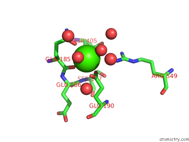
Mono view
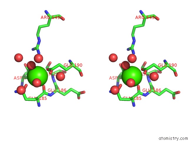
Stereo pair view
Mono view
Stereo pair view
A full contact list of Calcium with other atoms in the Ca binding
site number 1 of X-Tal Structure of the Mutant E44Q of the Cellulase CEL48F in Complex with A Thiooligosaccharide within 5.0Å range:
|
Calcium binding site 2 out of 2 in 1g9j
Go back to
Calcium binding site 2 out
of 2 in the X-Tal Structure of the Mutant E44Q of the Cellulase CEL48F in Complex with A Thiooligosaccharide
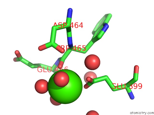
Mono view
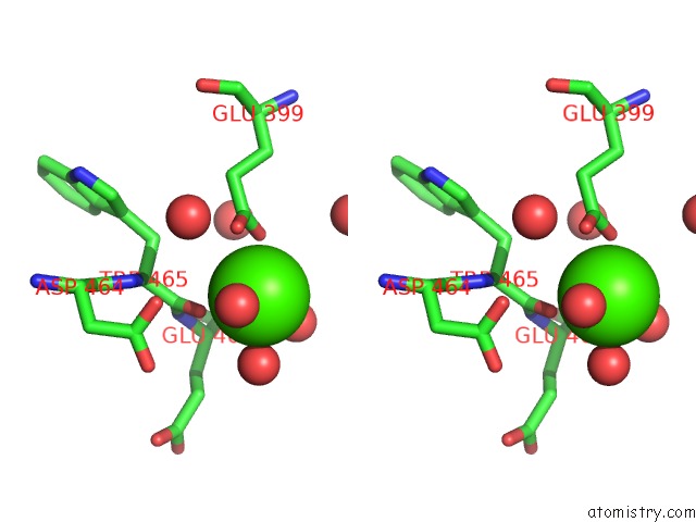
Stereo pair view
Mono view
Stereo pair view
A full contact list of Calcium with other atoms in the Ca binding
site number 2 of X-Tal Structure of the Mutant E44Q of the Cellulase CEL48F in Complex with A Thiooligosaccharide within 5.0Å range:
|
Reference:
G.Parsiegla,
C.Reverbel,
C.Tardif,
H.Driguez,
R.Haser.
Structures of Mutants of Cellulase CEL48F of Clostridium Cellulolyticum in Complex with Long Hemithiocellooligosaccharides Give Rise to A New View of the Substrate Pathway During Processive Action J.Mol.Biol. V. 375 499 2008.
ISSN: ISSN 0022-2836
PubMed: 18035374
DOI: 10.1016/J.JMB.2007.10.039
Page generated: Thu Jul 11 08:36:21 2024
ISSN: ISSN 0022-2836
PubMed: 18035374
DOI: 10.1016/J.JMB.2007.10.039
Last articles
Zn in 9J0NZn in 9J0O
Zn in 9J0P
Zn in 9FJX
Zn in 9EKB
Zn in 9C0F
Zn in 9CAH
Zn in 9CH0
Zn in 9CH3
Zn in 9CH1