Calcium »
PDB 1g8g-1gj6 »
1g9u »
Calcium in PDB 1g9u: Crystal Structure of Yopm-Leucine Rich Effector Protein From Yersinia Pestis
Protein crystallography data
The structure of Crystal Structure of Yopm-Leucine Rich Effector Protein From Yersinia Pestis, PDB code: 1g9u
was solved by
A.G.Evdokimov,
D.E.Anderson,
K.M.Routzahn,
D.S.Waugh,
with X-Ray Crystallography technique. A brief refinement statistics is given in the table below:
| Resolution Low / High (Å) | 100.00 / 2.35 |
| Space group | P 42 2 2 |
| Cell size a, b, c (Å), α, β, γ (°) | 109.359, 109.359, 101.499, 90.00, 90.00, 90.00 |
| R / Rfree (%) | 17 / 23 |
Other elements in 1g9u:
The structure of Crystal Structure of Yopm-Leucine Rich Effector Protein From Yersinia Pestis also contains other interesting chemical elements:
| Mercury | (Hg) | 8 atoms |
Calcium Binding Sites:
The binding sites of Calcium atom in the Crystal Structure of Yopm-Leucine Rich Effector Protein From Yersinia Pestis
(pdb code 1g9u). This binding sites where shown within
5.0 Angstroms radius around Calcium atom.
In total 2 binding sites of Calcium where determined in the Crystal Structure of Yopm-Leucine Rich Effector Protein From Yersinia Pestis, PDB code: 1g9u:
Jump to Calcium binding site number: 1; 2;
In total 2 binding sites of Calcium where determined in the Crystal Structure of Yopm-Leucine Rich Effector Protein From Yersinia Pestis, PDB code: 1g9u:
Jump to Calcium binding site number: 1; 2;
Calcium binding site 1 out of 2 in 1g9u
Go back to
Calcium binding site 1 out
of 2 in the Crystal Structure of Yopm-Leucine Rich Effector Protein From Yersinia Pestis
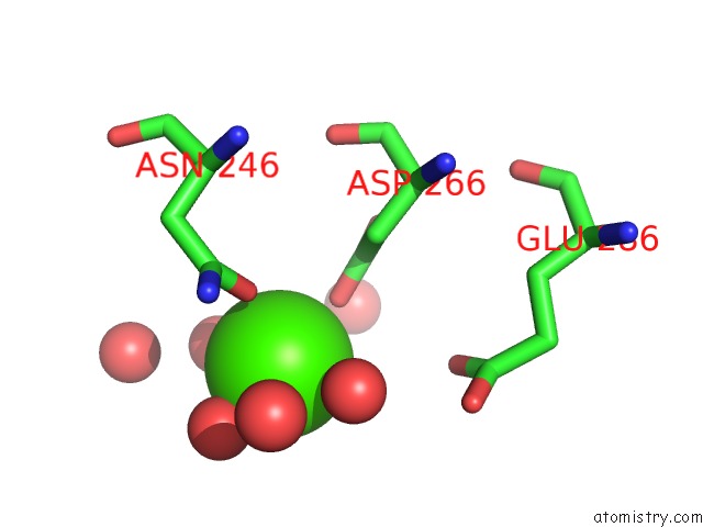
Mono view
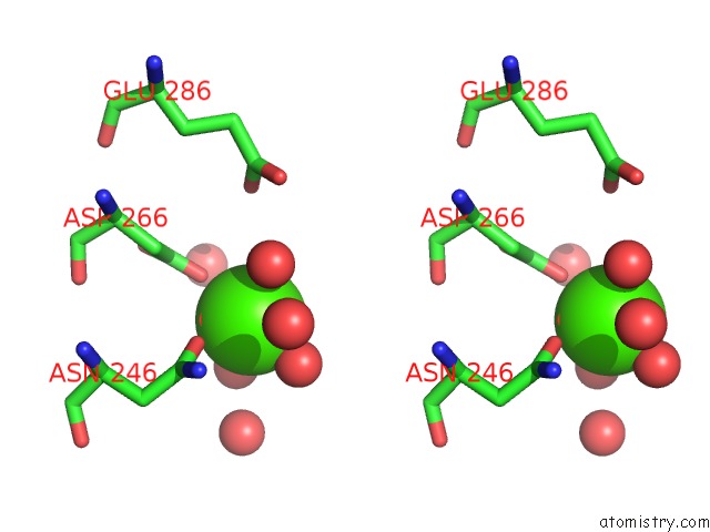
Stereo pair view
Mono view
Stereo pair view
A full contact list of Calcium with other atoms in the Ca binding
site number 1 of Crystal Structure of Yopm-Leucine Rich Effector Protein From Yersinia Pestis within 5.0Å range:
|
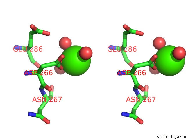
Calcium binding site 2 out of 2 in 1g9u
Go back to
Calcium binding site 2 out
of 2 in the Crystal Structure of Yopm-Leucine Rich Effector Protein From Yersinia Pestis
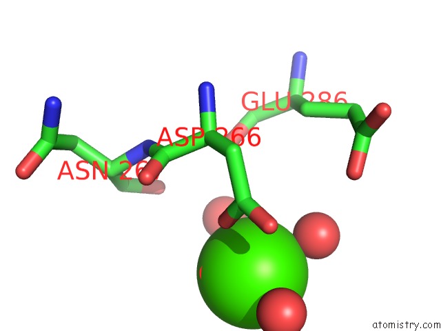
Mono view
Stereo pair view
Mono view
Stereo pair view
A full contact list of Calcium with other atoms in the Ca binding
site number 2 of Crystal Structure of Yopm-Leucine Rich Effector Protein From Yersinia Pestis within 5.0Å range:
|
Reference:
A.G.Evdokimov,
D.E.Anderson,
K.M.Routzahn,
D.S.Waugh.
Unusual Molecular Architecture of the Yersinia Pestis Cytotoxin Yopm: A Leucine-Rich Repeat Protein with the Shortest Repeating Unit. J.Mol.Biol. V. 312 807 2001.
ISSN: ISSN 0022-2836
PubMed: 11575934
DOI: 10.1006/JMBI.2001.4973
Page generated: Thu Jul 11 08:37:04 2024
ISSN: ISSN 0022-2836
PubMed: 11575934
DOI: 10.1006/JMBI.2001.4973
Last articles
Zn in 9MJ5Zn in 9HNW
Zn in 9G0L
Zn in 9FNE
Zn in 9DZN
Zn in 9E0I
Zn in 9D32
Zn in 9DAK
Zn in 8ZXC
Zn in 8ZUF