Calcium »
PDB 1g8g-1gj6 »
1gh4 »
Calcium in PDB 1gh4: Structure of the Triple Mutant (K56M, K120M, K121M) of Phospholipase A2
Enzymatic activity of Structure of the Triple Mutant (K56M, K120M, K121M) of Phospholipase A2
All present enzymatic activity of Structure of the Triple Mutant (K56M, K120M, K121M) of Phospholipase A2:
3.1.1.4;
3.1.1.4;
Protein crystallography data
The structure of Structure of the Triple Mutant (K56M, K120M, K121M) of Phospholipase A2, PDB code: 1gh4
was solved by
K.Sekar,
D.Velmurugan,
M.D.Tsai,
with X-Ray Crystallography technique. A brief refinement statistics is given in the table below:
| Resolution Low / High (Å) | 14.20 / 1.90 |
| Space group | P 1 2 1 |
| Cell size a, b, c (Å), α, β, γ (°) | 39.100, 24.350, 67.110, 90.00, 102.15, 90.00 |
| R / Rfree (%) | 19.6 / 25.9 |
Calcium Binding Sites:
The binding sites of Calcium atom in the Structure of the Triple Mutant (K56M, K120M, K121M) of Phospholipase A2
(pdb code 1gh4). This binding sites where shown within
5.0 Angstroms radius around Calcium atom.
In total 2 binding sites of Calcium where determined in the Structure of the Triple Mutant (K56M, K120M, K121M) of Phospholipase A2, PDB code: 1gh4:
Jump to Calcium binding site number: 1; 2;
In total 2 binding sites of Calcium where determined in the Structure of the Triple Mutant (K56M, K120M, K121M) of Phospholipase A2, PDB code: 1gh4:
Jump to Calcium binding site number: 1; 2;
Calcium binding site 1 out of 2 in 1gh4
Go back to
Calcium binding site 1 out
of 2 in the Structure of the Triple Mutant (K56M, K120M, K121M) of Phospholipase A2
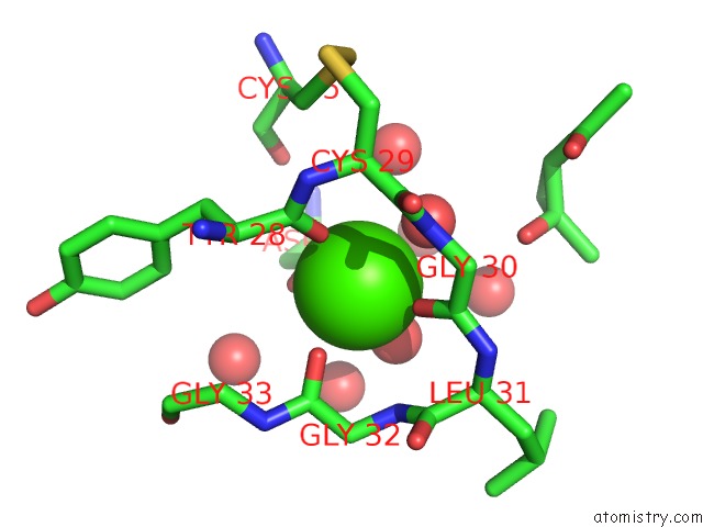
Mono view
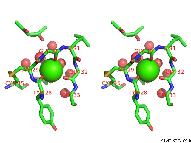
Stereo pair view
Mono view
Stereo pair view
A full contact list of Calcium with other atoms in the Ca binding
site number 1 of Structure of the Triple Mutant (K56M, K120M, K121M) of Phospholipase A2 within 5.0Å range:
|
Calcium binding site 2 out of 2 in 1gh4
Go back to
Calcium binding site 2 out
of 2 in the Structure of the Triple Mutant (K56M, K120M, K121M) of Phospholipase A2
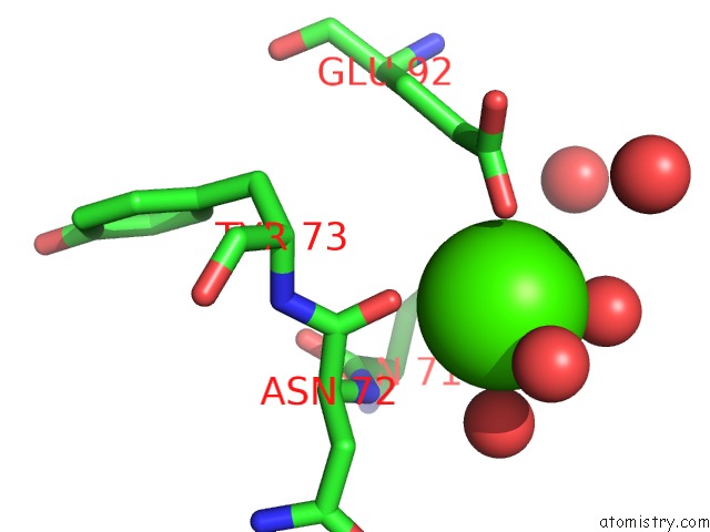
Mono view
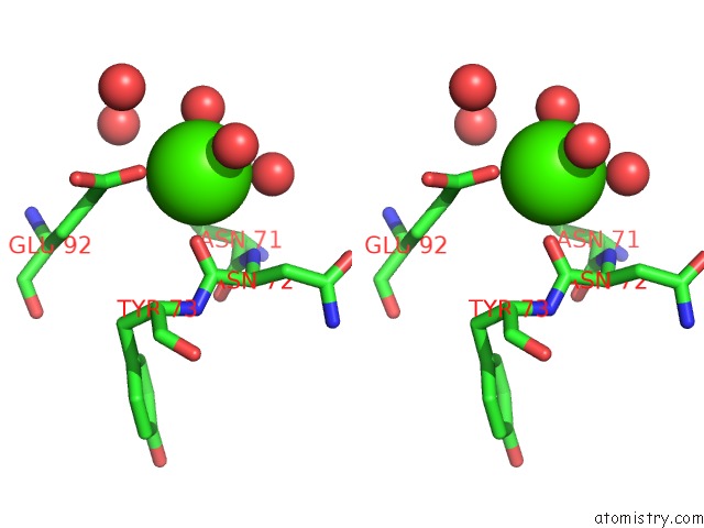
Stereo pair view
Mono view
Stereo pair view
A full contact list of Calcium with other atoms in the Ca binding
site number 2 of Structure of the Triple Mutant (K56M, K120M, K121M) of Phospholipase A2 within 5.0Å range:
|
Reference:
V.Rajakannan,
M.Yogavel,
M.J.Poi,
A.A.Jeyaprakash,
J.Jeyakanthan,
D.Velmurugan,
M.D.Tsai,
K.Sekar.
Observation of Additional Calcium Ion in the Crystal Structure of the Triple Mutant K56,120,121M of Bovine Pancreatic Phospholipase A2. J.Mol.Biol. V. 324 755 2002.
ISSN: ISSN 0022-2836
PubMed: 12460575
DOI: 10.1016/S0022-2836(02)01132-4
Page generated: Thu Jul 11 08:41:05 2024
ISSN: ISSN 0022-2836
PubMed: 12460575
DOI: 10.1016/S0022-2836(02)01132-4
Last articles
Zn in 9MJ5Zn in 9HNW
Zn in 9G0L
Zn in 9FNE
Zn in 9DZN
Zn in 9E0I
Zn in 9D32
Zn in 9DAK
Zn in 8ZXC
Zn in 8ZUF