Calcium »
PDB 1gk9-1gxo »
1gtt »
Calcium in PDB 1gtt: Crystal Structure of Hpce
Enzymatic activity of Crystal Structure of Hpce
All present enzymatic activity of Crystal Structure of Hpce:
5.3.3.10;
5.3.3.10;
Protein crystallography data
The structure of Crystal Structure of Hpce, PDB code: 1gtt
was solved by
J.R.H.Tame,
K.Namba,
E.J.Dodson,
D.I.Roper,
with X-Ray Crystallography technique. A brief refinement statistics is given in the table below:
| Resolution Low / High (Å) | 91.29 / 1.70 |
| Space group | P 21 21 21 |
| Cell size a, b, c (Å), α, β, γ (°) | 126.084, 138.201, 103.973, 90.00, 90.00, 90.00 |
| R / Rfree (%) | 21.6 / 25.2 |
Calcium Binding Sites:
The binding sites of Calcium atom in the Crystal Structure of Hpce
(pdb code 1gtt). This binding sites where shown within
5.0 Angstroms radius around Calcium atom.
In total 4 binding sites of Calcium where determined in the Crystal Structure of Hpce, PDB code: 1gtt:
Jump to Calcium binding site number: 1; 2; 3; 4;
In total 4 binding sites of Calcium where determined in the Crystal Structure of Hpce, PDB code: 1gtt:
Jump to Calcium binding site number: 1; 2; 3; 4;
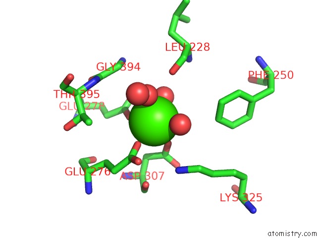
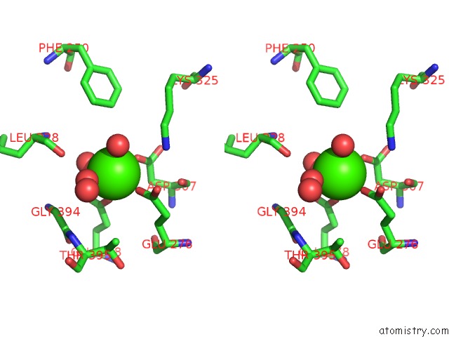
Calcium binding site 1 out of 4 in 1gtt
Go back to
Calcium binding site 1 out
of 4 in the Crystal Structure of Hpce
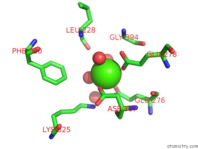
Mono view
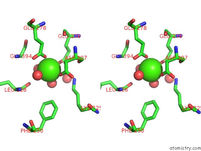
Stereo pair view
Mono view
Stereo pair view
A full contact list of Calcium with other atoms in the Ca binding
site number 1 of Crystal Structure of Hpce within 5.0Å range:
|
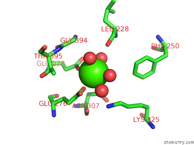
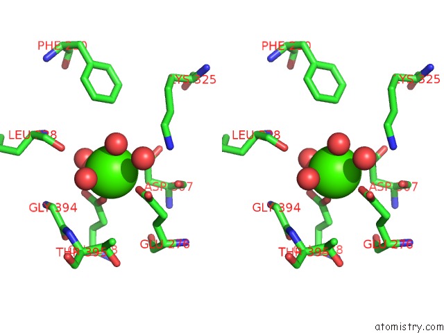
Calcium binding site 2 out of 4 in 1gtt
Go back to
Calcium binding site 2 out
of 4 in the Crystal Structure of Hpce
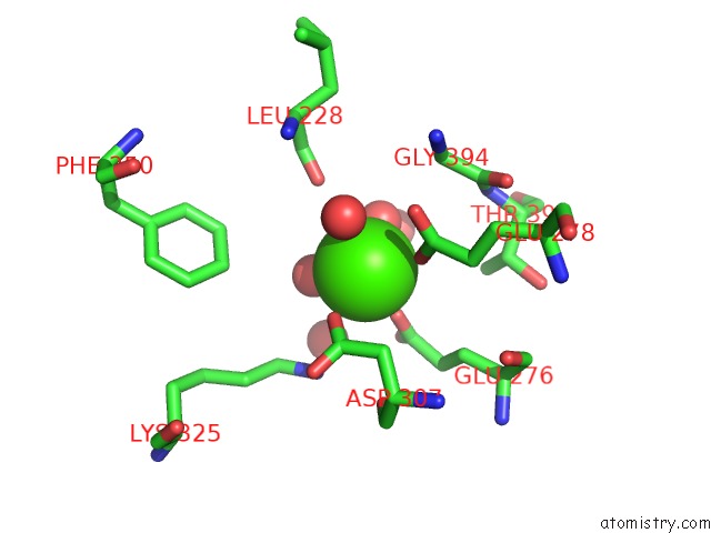
Mono view
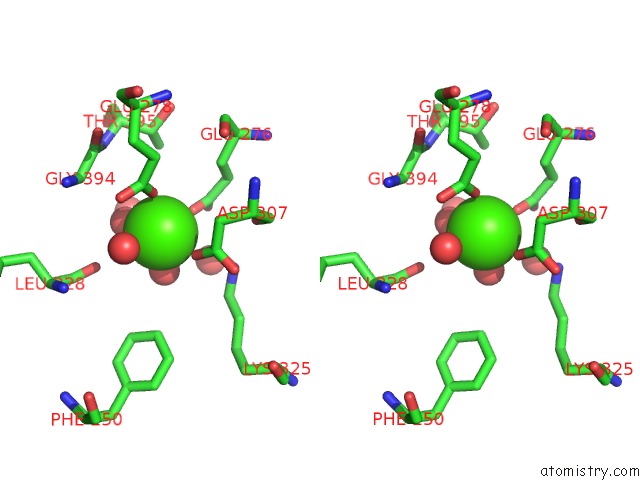
Stereo pair view
Mono view
Stereo pair view
A full contact list of Calcium with other atoms in the Ca binding
site number 2 of Crystal Structure of Hpce within 5.0Å range:
|
Calcium binding site 3 out of 4 in 1gtt
Go back to
Calcium binding site 3 out
of 4 in the Crystal Structure of Hpce
Mono view
Stereo pair view
Mono view
Stereo pair view
A full contact list of Calcium with other atoms in the Ca binding
site number 3 of Crystal Structure of Hpce within 5.0Å range:
|
Calcium binding site 4 out of 4 in 1gtt
Go back to
Calcium binding site 4 out
of 4 in the Crystal Structure of Hpce
Mono view
Stereo pair view
Mono view
Stereo pair view
A full contact list of Calcium with other atoms in the Ca binding
site number 4 of Crystal Structure of Hpce within 5.0Å range:
|
Reference:
J.R.H.Tame,
K.Namba,
E.J.Dodson,
D.I.Roper.
The Crystal Structure of Hpce, A Bifunctional Decarboxylase/Isomerase with A Multifunctional Fold. Biochemistry V. 41 2982 2002.
ISSN: ISSN 0006-2960
PubMed: 11863436
DOI: 10.1021/BI015717T
Page generated: Thu Jul 11 08:50:12 2024
ISSN: ISSN 0006-2960
PubMed: 11863436
DOI: 10.1021/BI015717T
Last articles
Zn in 9J0NZn in 9J0O
Zn in 9J0P
Zn in 9FJX
Zn in 9EKB
Zn in 9C0F
Zn in 9CAH
Zn in 9CH0
Zn in 9CH3
Zn in 9CH1