Calcium »
PDB 1gk9-1gxo »
1gw9 »
Calcium in PDB 1gw9: Tri-Iodide Derivative of Xylose Isomerase From Streptomyces Rubiginosus
Enzymatic activity of Tri-Iodide Derivative of Xylose Isomerase From Streptomyces Rubiginosus
All present enzymatic activity of Tri-Iodide Derivative of Xylose Isomerase From Streptomyces Rubiginosus:
5.3.1.5;
5.3.1.5;
Protein crystallography data
The structure of Tri-Iodide Derivative of Xylose Isomerase From Streptomyces Rubiginosus, PDB code: 1gw9
was solved by
G.Evans,
G.Bricogne,
with X-Ray Crystallography technique. A brief refinement statistics is given in the table below:
| Resolution Low / High (Å) | 32.05 / 1.55 |
| Space group | I 2 2 2 |
| Cell size a, b, c (Å), α, β, γ (°) | 92.354, 98.540, 102.507, 90.00, 90.00, 90.00 |
| R / Rfree (%) | 16.4 / 18.8 |
Other elements in 1gw9:
The structure of Tri-Iodide Derivative of Xylose Isomerase From Streptomyces Rubiginosus also contains other interesting chemical elements:
| Iodine | (I) | 29 atoms |
Calcium Binding Sites:
The binding sites of Calcium atom in the Tri-Iodide Derivative of Xylose Isomerase From Streptomyces Rubiginosus
(pdb code 1gw9). This binding sites where shown within
5.0 Angstroms radius around Calcium atom.
In total 2 binding sites of Calcium where determined in the Tri-Iodide Derivative of Xylose Isomerase From Streptomyces Rubiginosus, PDB code: 1gw9:
Jump to Calcium binding site number: 1; 2;
In total 2 binding sites of Calcium where determined in the Tri-Iodide Derivative of Xylose Isomerase From Streptomyces Rubiginosus, PDB code: 1gw9:
Jump to Calcium binding site number: 1; 2;
Calcium binding site 1 out of 2 in 1gw9
Go back to
Calcium binding site 1 out
of 2 in the Tri-Iodide Derivative of Xylose Isomerase From Streptomyces Rubiginosus
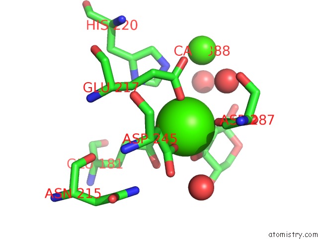
Mono view
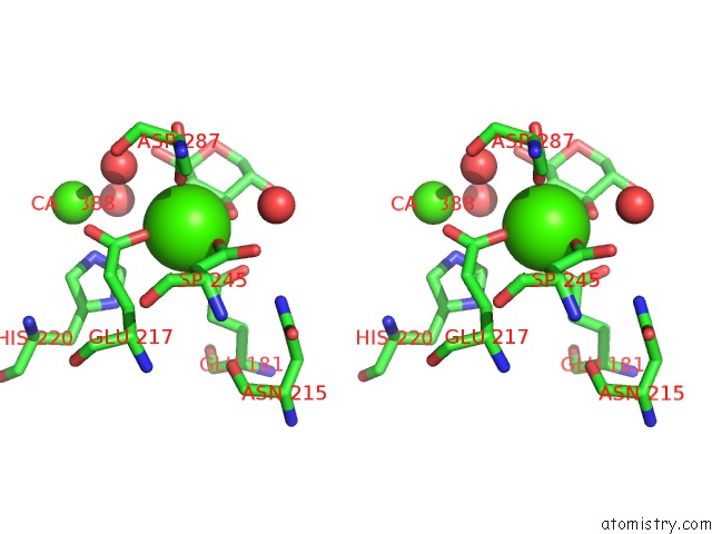
Stereo pair view
Mono view
Stereo pair view
A full contact list of Calcium with other atoms in the Ca binding
site number 1 of Tri-Iodide Derivative of Xylose Isomerase From Streptomyces Rubiginosus within 5.0Å range:
|
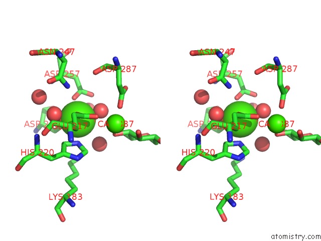
Calcium binding site 2 out of 2 in 1gw9
Go back to
Calcium binding site 2 out
of 2 in the Tri-Iodide Derivative of Xylose Isomerase From Streptomyces Rubiginosus
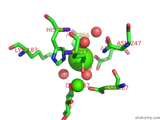
Mono view
Stereo pair view
Mono view
Stereo pair view
A full contact list of Calcium with other atoms in the Ca binding
site number 2 of Tri-Iodide Derivative of Xylose Isomerase From Streptomyces Rubiginosus within 5.0Å range:
|
Reference:
G.Evans,
G.Bricogne.
Triiodide Derivatization and Combinatorial Counter-Ion Replacement: Two Methods For Enhancing Phasing Signal Using Laboratory Cu Kalpha X-Ray Equipment Acta Crystallogr.,Sect.D V. 58 976 2002.
ISSN: ISSN 0907-4449
PubMed: 12037300
DOI: 10.1107/S0907444902005486
Page generated: Mon Jul 7 15:22:53 2025
ISSN: ISSN 0907-4449
PubMed: 12037300
DOI: 10.1107/S0907444902005486
Last articles
Cl in 8E1WCl in 8E1Z
Cl in 8E1F
Cl in 8DZS
Cl in 8DZR
Cl in 8DZ1
Cl in 8DZ7
Cl in 8DYZ
Cl in 8DZ0
Cl in 8DYH