Calcium »
PDB 1h80-1hny »
1hm0 »
Calcium in PDB 1hm0: Crystal Structure of S.Pneumoniae N-Acetylglucosamine 1-Phosphate Uridyltransferase, Glmu
Enzymatic activity of Crystal Structure of S.Pneumoniae N-Acetylglucosamine 1-Phosphate Uridyltransferase, Glmu
All present enzymatic activity of Crystal Structure of S.Pneumoniae N-Acetylglucosamine 1-Phosphate Uridyltransferase, Glmu:
2.7.7.23;
2.7.7.23;
Protein crystallography data
The structure of Crystal Structure of S.Pneumoniae N-Acetylglucosamine 1-Phosphate Uridyltransferase, Glmu, PDB code: 1hm0
was solved by
G.Sulzenbacher,
L.Gal,
C.Peneff,
F.Fassy,
Y.Bourne,
with X-Ray Crystallography technique. A brief refinement statistics is given in the table below:
| Resolution Low / High (Å) | 40.00 / 2.30 |
| Space group | H 3 |
| Cell size a, b, c (Å), α, β, γ (°) | 92.715, 92.715, 280.387, 90.00, 90.00, 120.00 |
| R / Rfree (%) | 20.7 / 24.4 |
Calcium Binding Sites:
The binding sites of Calcium atom in the Crystal Structure of S.Pneumoniae N-Acetylglucosamine 1-Phosphate Uridyltransferase, Glmu
(pdb code 1hm0). This binding sites where shown within
5.0 Angstroms radius around Calcium atom.
In total 6 binding sites of Calcium where determined in the Crystal Structure of S.Pneumoniae N-Acetylglucosamine 1-Phosphate Uridyltransferase, Glmu, PDB code: 1hm0:
Jump to Calcium binding site number: 1; 2; 3; 4; 5; 6;
In total 6 binding sites of Calcium where determined in the Crystal Structure of S.Pneumoniae N-Acetylglucosamine 1-Phosphate Uridyltransferase, Glmu, PDB code: 1hm0:
Jump to Calcium binding site number: 1; 2; 3; 4; 5; 6;
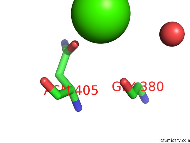
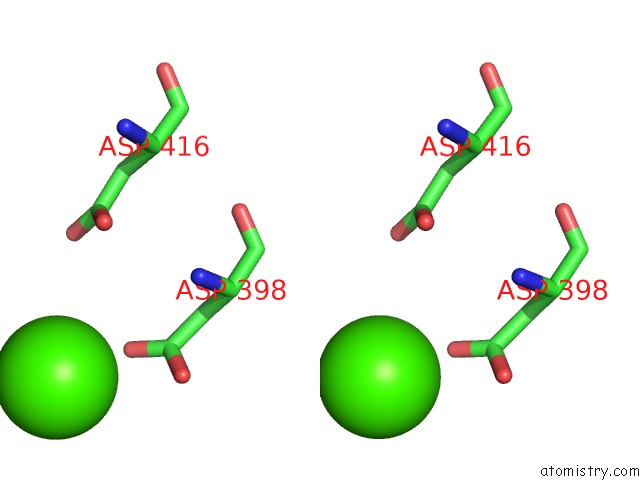
Calcium binding site 1 out of 6 in 1hm0
Go back to
Calcium binding site 1 out
of 6 in the Crystal Structure of S.Pneumoniae N-Acetylglucosamine 1-Phosphate Uridyltransferase, Glmu
Mono view
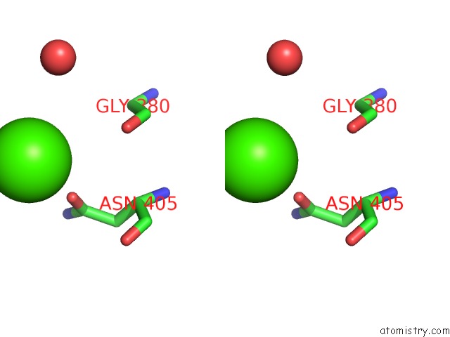
Stereo pair view
Mono view
Stereo pair view
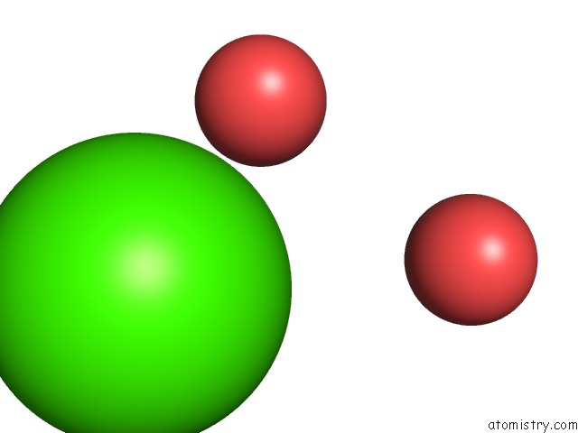
A full contact list of Calcium with other atoms in the Ca binding
site number 1 of Crystal Structure of S.Pneumoniae N-Acetylglucosamine 1-Phosphate Uridyltransferase, Glmu within 5.0Å range:
|
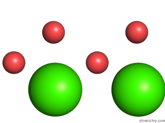
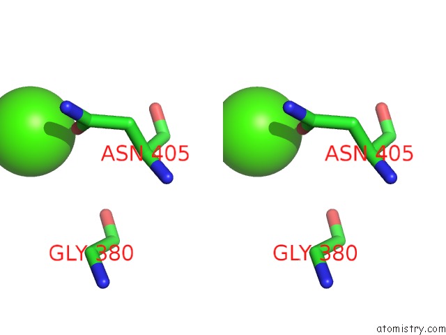
Calcium binding site 2 out of 6 in 1hm0
Go back to
Calcium binding site 2 out
of 6 in the Crystal Structure of S.Pneumoniae N-Acetylglucosamine 1-Phosphate Uridyltransferase, Glmu

Mono view

Stereo pair view
Mono view
Stereo pair view
A full contact list of Calcium with other atoms in the Ca binding
site number 2 of Crystal Structure of S.Pneumoniae N-Acetylglucosamine 1-Phosphate Uridyltransferase, Glmu within 5.0Å range:
|
Calcium binding site 3 out of 6 in 1hm0
Go back to
Calcium binding site 3 out
of 6 in the Crystal Structure of S.Pneumoniae N-Acetylglucosamine 1-Phosphate Uridyltransferase, Glmu

Mono view
Stereo pair view
Mono view
Stereo pair view
A full contact list of Calcium with other atoms in the Ca binding
site number 3 of Crystal Structure of S.Pneumoniae N-Acetylglucosamine 1-Phosphate Uridyltransferase, Glmu within 5.0Å range:
|
Calcium binding site 4 out of 6 in 1hm0
Go back to
Calcium binding site 4 out
of 6 in the Crystal Structure of S.Pneumoniae N-Acetylglucosamine 1-Phosphate Uridyltransferase, Glmu

Mono view
Stereo pair view
Mono view
Stereo pair view
A full contact list of Calcium with other atoms in the Ca binding
site number 4 of Crystal Structure of S.Pneumoniae N-Acetylglucosamine 1-Phosphate Uridyltransferase, Glmu within 5.0Å range:
|
Calcium binding site 5 out of 6 in 1hm0
Go back to
Calcium binding site 5 out
of 6 in the Crystal Structure of S.Pneumoniae N-Acetylglucosamine 1-Phosphate Uridyltransferase, Glmu

Mono view
Stereo pair view
Mono view
Stereo pair view
A full contact list of Calcium with other atoms in the Ca binding
site number 5 of Crystal Structure of S.Pneumoniae N-Acetylglucosamine 1-Phosphate Uridyltransferase, Glmu within 5.0Å range:
|
Calcium binding site 6 out of 6 in 1hm0
Go back to
Calcium binding site 6 out
of 6 in the Crystal Structure of S.Pneumoniae N-Acetylglucosamine 1-Phosphate Uridyltransferase, Glmu
Mono view

Stereo pair view
Mono view
Stereo pair view
A full contact list of Calcium with other atoms in the Ca binding
site number 6 of Crystal Structure of S.Pneumoniae N-Acetylglucosamine 1-Phosphate Uridyltransferase, Glmu within 5.0Å range:
|
Reference:
G.Sulzenbacher,
L.Gal,
C.Peneff,
F.Fassy,
Y.Bourne.
Crystal Structure of Streptococcus Pneumoniae N-Acetylglucosamine-1-Phosphate Uridyltransferase Bound to Acetyl-Coenzyme A Reveals A Novel Active Site Architecture. J.Biol.Chem. V. 276 11844 2001.
ISSN: ISSN 0021-9258
PubMed: 11118459
DOI: 10.1074/JBC.M011225200
Page generated: Thu Jul 11 10:04:28 2024
ISSN: ISSN 0021-9258
PubMed: 11118459
DOI: 10.1074/JBC.M011225200
Last articles
Zn in 9J0NZn in 9J0O
Zn in 9J0P
Zn in 9FJX
Zn in 9EKB
Zn in 9C0F
Zn in 9CAH
Zn in 9CH0
Zn in 9CH3
Zn in 9CH1