Calcium »
PDB 1h80-1hny »
1hn4 »
Calcium in PDB 1hn4: Prophospholipase A2 Dimer Complexed with MJ33, Sulfate, and Calcium
Enzymatic activity of Prophospholipase A2 Dimer Complexed with MJ33, Sulfate, and Calcium
All present enzymatic activity of Prophospholipase A2 Dimer Complexed with MJ33, Sulfate, and Calcium:
3.1.1.4;
3.1.1.4;
Protein crystallography data
The structure of Prophospholipase A2 Dimer Complexed with MJ33, Sulfate, and Calcium, PDB code: 1hn4
was solved by
T.M.Epstein,
Y.H.Pan,
M.K.Jain,
B.J.Bahnson,
with X-Ray Crystallography technique. A brief refinement statistics is given in the table below:
| Resolution Low / High (Å) | 20.00 / 1.50 |
| Space group | P 1 21 1 |
| Cell size a, b, c (Å), α, β, γ (°) | 37.488, 54.852, 56.896, 90.00, 104.06, 90.00 |
| R / Rfree (%) | 21.5 / 23.9 |
Other elements in 1hn4:
The structure of Prophospholipase A2 Dimer Complexed with MJ33, Sulfate, and Calcium also contains other interesting chemical elements:
| Fluorine | (F) | 3 atoms |
Calcium Binding Sites:
The binding sites of Calcium atom in the Prophospholipase A2 Dimer Complexed with MJ33, Sulfate, and Calcium
(pdb code 1hn4). This binding sites where shown within
5.0 Angstroms radius around Calcium atom.
In total 2 binding sites of Calcium where determined in the Prophospholipase A2 Dimer Complexed with MJ33, Sulfate, and Calcium, PDB code: 1hn4:
Jump to Calcium binding site number: 1; 2;
In total 2 binding sites of Calcium where determined in the Prophospholipase A2 Dimer Complexed with MJ33, Sulfate, and Calcium, PDB code: 1hn4:
Jump to Calcium binding site number: 1; 2;
Calcium binding site 1 out of 2 in 1hn4
Go back to
Calcium binding site 1 out
of 2 in the Prophospholipase A2 Dimer Complexed with MJ33, Sulfate, and Calcium
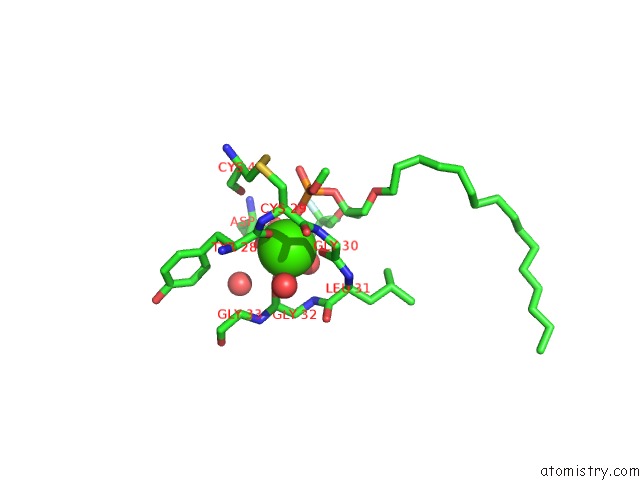
Mono view
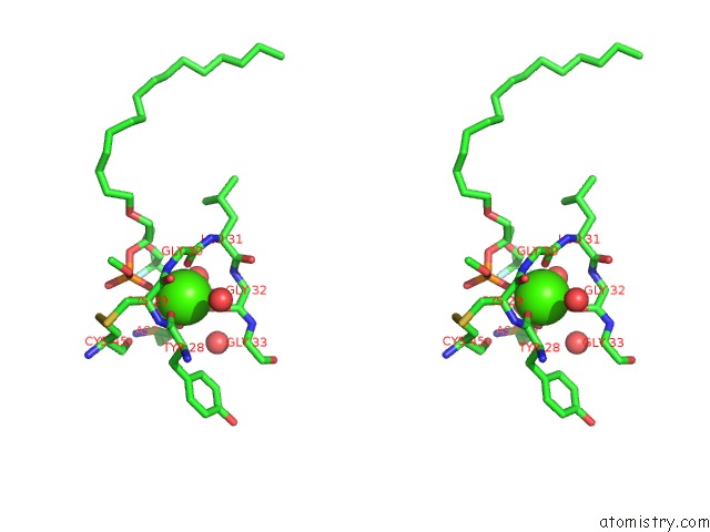
Stereo pair view
Mono view
Stereo pair view
A full contact list of Calcium with other atoms in the Ca binding
site number 1 of Prophospholipase A2 Dimer Complexed with MJ33, Sulfate, and Calcium within 5.0Å range:
|
Calcium binding site 2 out of 2 in 1hn4
Go back to
Calcium binding site 2 out
of 2 in the Prophospholipase A2 Dimer Complexed with MJ33, Sulfate, and Calcium
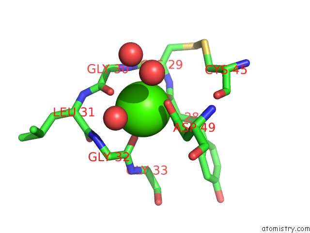
Mono view
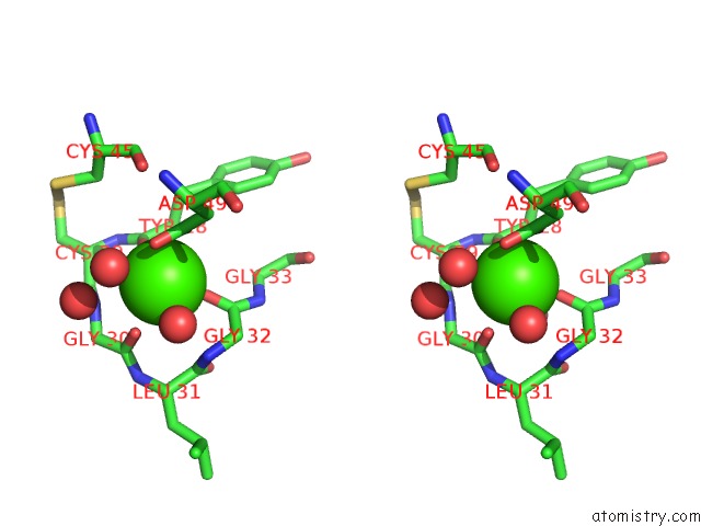
Stereo pair view
Mono view
Stereo pair view
A full contact list of Calcium with other atoms in the Ca binding
site number 2 of Prophospholipase A2 Dimer Complexed with MJ33, Sulfate, and Calcium within 5.0Å range:
|
Reference:
T.M.Epstein,
B.Z.Yu,
Y.H.Pan,
S.P.Tutton,
B.P.Maliwal,
M.K.Jain,
B.J.Bahnson.
The Basis For K(Cat) Impairment in Prophospholipase A(2) From the Anion-Assisted Dimer Structure. Biochemistry V. 40 11411 2001.
ISSN: ISSN 0006-2960
PubMed: 11560489
DOI: 10.1021/BI011228H
Page generated: Mon Jul 7 15:39:42 2025
ISSN: ISSN 0006-2960
PubMed: 11560489
DOI: 10.1021/BI011228H
Last articles
F in 7PY4F in 7PX6
F in 7PVU
F in 7PRV
F in 7PRW
F in 7PMQ
F in 7PRX
F in 7PPH
F in 7PRM
F in 7PQV