Calcium »
PDB 1hov-1i6t »
1hvd »
Calcium in PDB 1hvd: Structural and Electrophysiological Analysis of Annexin V Mutants. Mutagenesis of Human Annexin V, An in Vitro Voltage-Gated Calcium Channel, Provides Information About the Structural Features of the Ion Pathway, the Voltage Sensor and the Ion Selectivity Filter
Protein crystallography data
The structure of Structural and Electrophysiological Analysis of Annexin V Mutants. Mutagenesis of Human Annexin V, An in Vitro Voltage-Gated Calcium Channel, Provides Information About the Structural Features of the Ion Pathway, the Voltage Sensor and the Ion Selectivity Filter, PDB code: 1hvd
was solved by
A.Burger,
R.Huber,
with X-Ray Crystallography technique. A brief refinement statistics is given in the table below:
| Resolution Low / High (Å) | N/A / 2.00 |
| Space group | H 3 |
| Cell size a, b, c (Å), α, β, γ (°) | 99.600, 99.600, 96.600, 90.00, 90.00, 120.00 |
| R / Rfree (%) | 18.9 / n/a |
Calcium Binding Sites:
The binding sites of Calcium atom in the Structural and Electrophysiological Analysis of Annexin V Mutants. Mutagenesis of Human Annexin V, An in Vitro Voltage-Gated Calcium Channel, Provides Information About the Structural Features of the Ion Pathway, the Voltage Sensor and the Ion Selectivity Filter
(pdb code 1hvd). This binding sites where shown within
5.0 Angstroms radius around Calcium atom.
In total 4 binding sites of Calcium where determined in the Structural and Electrophysiological Analysis of Annexin V Mutants. Mutagenesis of Human Annexin V, An in Vitro Voltage-Gated Calcium Channel, Provides Information About the Structural Features of the Ion Pathway, the Voltage Sensor and the Ion Selectivity Filter, PDB code: 1hvd:
Jump to Calcium binding site number: 1; 2; 3; 4;
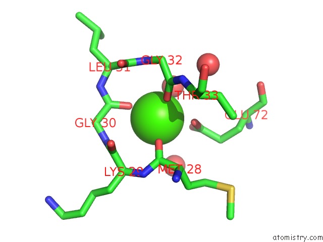
Calcium binding site 1 out of 4 in 1hvd
Go back to
Calcium binding site 1 out
of 4 in the Structural and Electrophysiological Analysis of Annexin V Mutants. Mutagenesis of Human Annexin V, An in Vitro Voltage-Gated Calcium Channel, Provides Information About the Structural Features of the Ion Pathway, the Voltage Sensor and the Ion Selectivity Filter
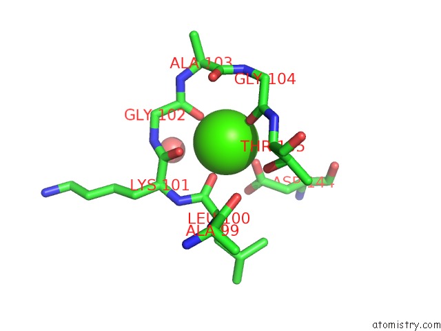
Mono view
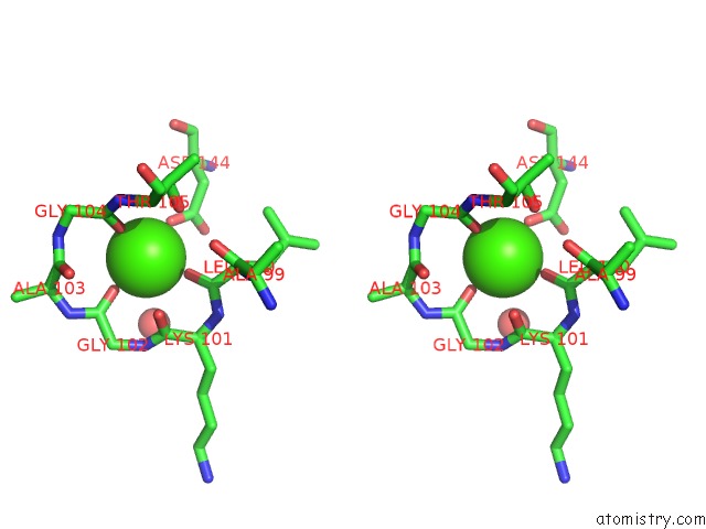
Stereo pair view
Mono view
Stereo pair view
A full contact list of Calcium with other atoms in the Ca binding
site number 1 of Structural and Electrophysiological Analysis of Annexin V Mutants. Mutagenesis of Human Annexin V, An in Vitro Voltage-Gated Calcium Channel, Provides Information About the Structural Features of the Ion Pathway, the Voltage Sensor and the Ion Selectivity Filter within 5.0Å range:
|
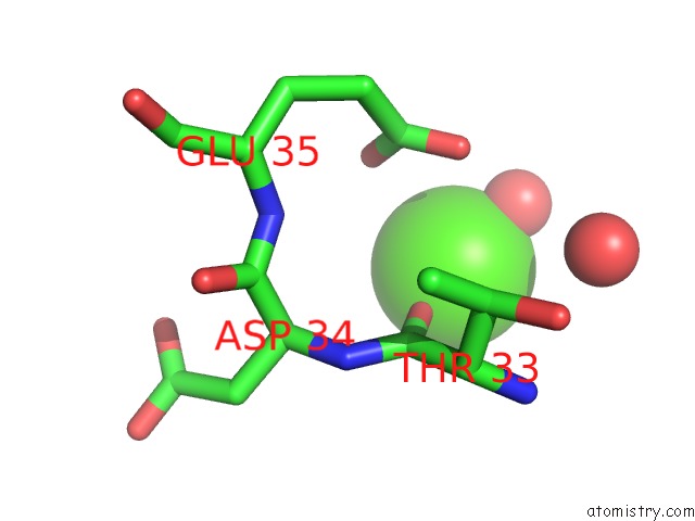
Calcium binding site 2 out of 4 in 1hvd
Go back to
Calcium binding site 2 out
of 4 in the Structural and Electrophysiological Analysis of Annexin V Mutants. Mutagenesis of Human Annexin V, An in Vitro Voltage-Gated Calcium Channel, Provides Information About the Structural Features of the Ion Pathway, the Voltage Sensor and the Ion Selectivity Filter
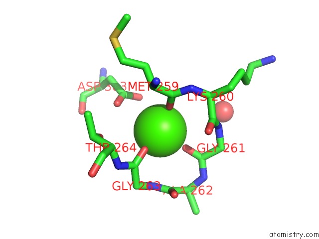
Mono view
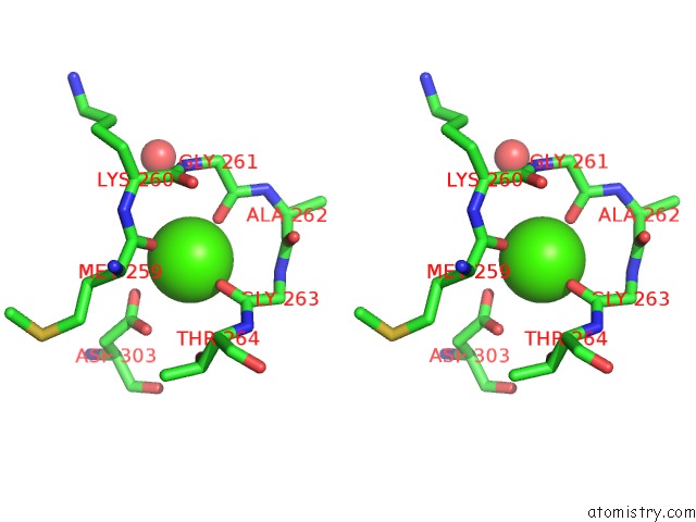
Stereo pair view
Mono view
Stereo pair view
A full contact list of Calcium with other atoms in the Ca binding
site number 2 of Structural and Electrophysiological Analysis of Annexin V Mutants. Mutagenesis of Human Annexin V, An in Vitro Voltage-Gated Calcium Channel, Provides Information About the Structural Features of the Ion Pathway, the Voltage Sensor and the Ion Selectivity Filter within 5.0Å range:
|
Calcium binding site 3 out of 4 in 1hvd
Go back to
Calcium binding site 3 out
of 4 in the Structural and Electrophysiological Analysis of Annexin V Mutants. Mutagenesis of Human Annexin V, An in Vitro Voltage-Gated Calcium Channel, Provides Information About the Structural Features of the Ion Pathway, the Voltage Sensor and the Ion Selectivity Filter
Mono view

Stereo pair view
Mono view
Stereo pair view
A full contact list of Calcium with other atoms in the Ca binding
site number 3 of Structural and Electrophysiological Analysis of Annexin V Mutants. Mutagenesis of Human Annexin V, An in Vitro Voltage-Gated Calcium Channel, Provides Information About the Structural Features of the Ion Pathway, the Voltage Sensor and the Ion Selectivity Filter within 5.0Å range:
|
Calcium binding site 4 out of 4 in 1hvd
Go back to
Calcium binding site 4 out
of 4 in the Structural and Electrophysiological Analysis of Annexin V Mutants. Mutagenesis of Human Annexin V, An in Vitro Voltage-Gated Calcium Channel, Provides Information About the Structural Features of the Ion Pathway, the Voltage Sensor and the Ion Selectivity Filter
Mono view

Stereo pair view
Mono view
Stereo pair view
A full contact list of Calcium with other atoms in the Ca binding
site number 4 of Structural and Electrophysiological Analysis of Annexin V Mutants. Mutagenesis of Human Annexin V, An in Vitro Voltage-Gated Calcium Channel, Provides Information About the Structural Features of the Ion Pathway, the Voltage Sensor and the Ion Selectivity Filter within 5.0Å range:
|
Reference:
A.Burger,
D.Voges,
P.Demange,
C.R.Perez,
R.Huber,
R.Berendes.
Structural and Electrophysiological Analysis of Annexin V Mutants. Mutagenesis of Human Annexin V, An in Vitro Voltage-Gated Calcium Channel, Provides Information About the Structural Features of the Ion Pathway, the Voltage Sensor and the Ion Selectivity Filter J.Mol.Biol. V. 237 479 1994.
ISSN: ISSN 0022-2836
PubMed: 8151707
DOI: 10.1006/JMBI.1994.1249
Page generated: Thu Jul 11 10:09:58 2024
ISSN: ISSN 0022-2836
PubMed: 8151707
DOI: 10.1006/JMBI.1994.1249
Last articles
Zn in 9J0NZn in 9J0O
Zn in 9J0P
Zn in 9FJX
Zn in 9EKB
Zn in 9C0F
Zn in 9CAH
Zn in 9CH0
Zn in 9CH3
Zn in 9CH1