Calcium »
PDB 1hov-1i6t »
1hyo »
Calcium in PDB 1hyo: Crystal Structure of Fumarylacetoacetate Hydrolase Complexed with 4-(Hydroxymethylphosphinoyl)-3-Oxo-Butanoic Acid
Enzymatic activity of Crystal Structure of Fumarylacetoacetate Hydrolase Complexed with 4-(Hydroxymethylphosphinoyl)-3-Oxo-Butanoic Acid
All present enzymatic activity of Crystal Structure of Fumarylacetoacetate Hydrolase Complexed with 4-(Hydroxymethylphosphinoyl)-3-Oxo-Butanoic Acid:
3.7.1.2;
3.7.1.2;
Protein crystallography data
The structure of Crystal Structure of Fumarylacetoacetate Hydrolase Complexed with 4-(Hydroxymethylphosphinoyl)-3-Oxo-Butanoic Acid, PDB code: 1hyo
was solved by
R.L.Bateman,
P.Bhanumoorthy,
J.F.Witte,
R.W.Mcclard,
M.Grompe,
D.E.Timm,
with X-Ray Crystallography technique. A brief refinement statistics is given in the table below:
| Resolution Low / High (Å) | 27.00 / 1.30 |
| Space group | P 1 21 1 |
| Cell size a, b, c (Å), α, β, γ (°) | 64.102, 109.458, 67.459, 90.00, 102.37, 90.00 |
| R / Rfree (%) | 18.1 / 19.9 |
Other elements in 1hyo:
The structure of Crystal Structure of Fumarylacetoacetate Hydrolase Complexed with 4-(Hydroxymethylphosphinoyl)-3-Oxo-Butanoic Acid also contains other interesting chemical elements:
| Nickel | (Ni) | 1 atom |
| Magnesium | (Mg) | 2 atoms |
Calcium Binding Sites:
The binding sites of Calcium atom in the Crystal Structure of Fumarylacetoacetate Hydrolase Complexed with 4-(Hydroxymethylphosphinoyl)-3-Oxo-Butanoic Acid
(pdb code 1hyo). This binding sites where shown within
5.0 Angstroms radius around Calcium atom.
In total 2 binding sites of Calcium where determined in the Crystal Structure of Fumarylacetoacetate Hydrolase Complexed with 4-(Hydroxymethylphosphinoyl)-3-Oxo-Butanoic Acid, PDB code: 1hyo:
Jump to Calcium binding site number: 1; 2;
In total 2 binding sites of Calcium where determined in the Crystal Structure of Fumarylacetoacetate Hydrolase Complexed with 4-(Hydroxymethylphosphinoyl)-3-Oxo-Butanoic Acid, PDB code: 1hyo:
Jump to Calcium binding site number: 1; 2;
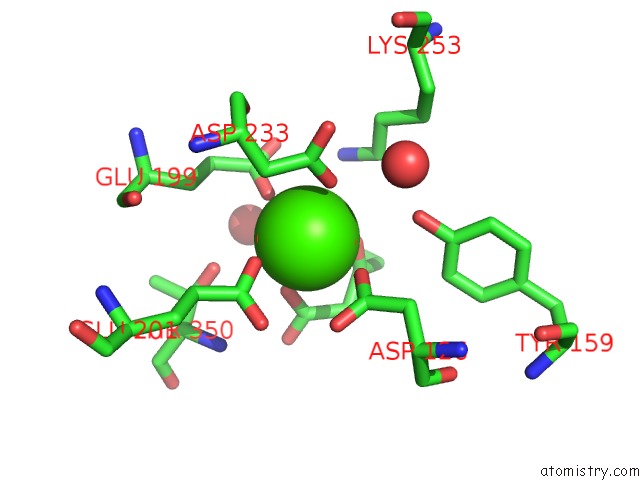
Calcium binding site 1 out of 2 in 1hyo
Go back to
Calcium binding site 1 out
of 2 in the Crystal Structure of Fumarylacetoacetate Hydrolase Complexed with 4-(Hydroxymethylphosphinoyl)-3-Oxo-Butanoic Acid
Mono view

Stereo pair view
Mono view
Stereo pair view
A full contact list of Calcium with other atoms in the Ca binding
site number 1 of Crystal Structure of Fumarylacetoacetate Hydrolase Complexed with 4-(Hydroxymethylphosphinoyl)-3-Oxo-Butanoic Acid within 5.0Å range:
|
Calcium binding site 2 out of 2 in 1hyo
Go back to
Calcium binding site 2 out
of 2 in the Crystal Structure of Fumarylacetoacetate Hydrolase Complexed with 4-(Hydroxymethylphosphinoyl)-3-Oxo-Butanoic Acid

Mono view

Stereo pair view
Mono view
Stereo pair view
A full contact list of Calcium with other atoms in the Ca binding
site number 2 of Crystal Structure of Fumarylacetoacetate Hydrolase Complexed with 4-(Hydroxymethylphosphinoyl)-3-Oxo-Butanoic Acid within 5.0Å range:
|
Reference:
R.L.Bateman,
P.Bhanumoorthy,
J.F.Witte,
R.W.Mcclard,
M.Grompe,
D.E.Timm.
Mechanistic Inferences From the Crystal Structure of Fumarylacetoacetate Hydrolase with A Bound Phosphorus-Based Inhibitor. J.Biol.Chem. V. 276 15284 2001.
ISSN: ISSN 0021-9258
PubMed: 11154690
DOI: 10.1074/JBC.M007621200
Page generated: Mon Jul 7 15:44:15 2025
ISSN: ISSN 0021-9258
PubMed: 11154690
DOI: 10.1074/JBC.M007621200
Last articles
Fe in 2YXOFe in 2YRS
Fe in 2YXC
Fe in 2YNM
Fe in 2YVJ
Fe in 2YP1
Fe in 2YU2
Fe in 2YU1
Fe in 2YQB
Fe in 2YOO