Calcium »
PDB 1hov-1i6t »
1i0x »
Calcium in PDB 1i0x: Ribonuclease T1 in Complex with 2'Gmp (Form II Crystal)
Enzymatic activity of Ribonuclease T1 in Complex with 2'Gmp (Form II Crystal)
All present enzymatic activity of Ribonuclease T1 in Complex with 2'Gmp (Form II Crystal):
3.1.27.3;
3.1.27.3;
Protein crystallography data
The structure of Ribonuclease T1 in Complex with 2'Gmp (Form II Crystal), PDB code: 1i0x
was solved by
J.De Swarte,
S.De Vos,
U.Langhorst,
J.Steyaert,
R.Loris,
with X-Ray Crystallography technique. A brief refinement statistics is given in the table below:
| Resolution Low / High (Å) | 15.00 / 1.65 |
| Space group | P 21 21 21 |
| Cell size a, b, c (Å), α, β, γ (°) | 58.271, 59.696, 100.136, 90.00, 90.00, 90.00 |
| R / Rfree (%) | 19 / 21.6 |
Calcium Binding Sites:
The binding sites of Calcium atom in the Ribonuclease T1 in Complex with 2'Gmp (Form II Crystal)
(pdb code 1i0x). This binding sites where shown within
5.0 Angstroms radius around Calcium atom.
In total 5 binding sites of Calcium where determined in the Ribonuclease T1 in Complex with 2'Gmp (Form II Crystal), PDB code: 1i0x:
Jump to Calcium binding site number: 1; 2; 3; 4; 5;
In total 5 binding sites of Calcium where determined in the Ribonuclease T1 in Complex with 2'Gmp (Form II Crystal), PDB code: 1i0x:
Jump to Calcium binding site number: 1; 2; 3; 4; 5;
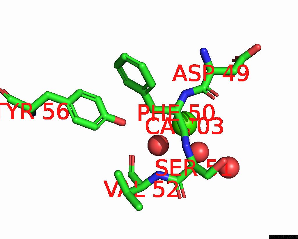
Calcium binding site 1 out of 5 in 1i0x
Go back to
Calcium binding site 1 out
of 5 in the Ribonuclease T1 in Complex with 2'Gmp (Form II Crystal)
Mono view

Stereo pair view
Mono view
Stereo pair view
A full contact list of Calcium with other atoms in the Ca binding
site number 1 of Ribonuclease T1 in Complex with 2'Gmp (Form II Crystal) within 5.0Å range:
|
Calcium binding site 2 out of 5 in 1i0x
Go back to
Calcium binding site 2 out
of 5 in the Ribonuclease T1 in Complex with 2'Gmp (Form II Crystal)

Mono view

Stereo pair view
Mono view
Stereo pair view
A full contact list of Calcium with other atoms in the Ca binding
site number 2 of Ribonuclease T1 in Complex with 2'Gmp (Form II Crystal) within 5.0Å range:
|
Calcium binding site 3 out of 5 in 1i0x
Go back to
Calcium binding site 3 out
of 5 in the Ribonuclease T1 in Complex with 2'Gmp (Form II Crystal)

Mono view

Stereo pair view
Mono view
Stereo pair view
A full contact list of Calcium with other atoms in the Ca binding
site number 3 of Ribonuclease T1 in Complex with 2'Gmp (Form II Crystal) within 5.0Å range:
|
Calcium binding site 4 out of 5 in 1i0x
Go back to
Calcium binding site 4 out
of 5 in the Ribonuclease T1 in Complex with 2'Gmp (Form II Crystal)

Mono view

Stereo pair view
Mono view
Stereo pair view
A full contact list of Calcium with other atoms in the Ca binding
site number 4 of Ribonuclease T1 in Complex with 2'Gmp (Form II Crystal) within 5.0Å range:
|
Calcium binding site 5 out of 5 in 1i0x
Go back to
Calcium binding site 5 out
of 5 in the Ribonuclease T1 in Complex with 2'Gmp (Form II Crystal)

Mono view

Stereo pair view
Mono view
Stereo pair view
A full contact list of Calcium with other atoms in the Ca binding
site number 5 of Ribonuclease T1 in Complex with 2'Gmp (Form II Crystal) within 5.0Å range:
|
Reference:
J.Deswarte,
S.De Vos,
U.Langhorst,
J.Steyaert,
R.Loris.
The Contribution of Metal Ions to the Conformational Stability of Ribonuclease T1: Crystal Versus Solution. Eur.J.Biochem. V. 268 3993 2001.
ISSN: ISSN 0014-2956
PubMed: 11453993
DOI: 10.1046/J.1432-1327.2001.02310.X
Page generated: Thu Jul 11 10:13:36 2024
ISSN: ISSN 0014-2956
PubMed: 11453993
DOI: 10.1046/J.1432-1327.2001.02310.X
Last articles
Zn in 9J0NZn in 9J0O
Zn in 9J0P
Zn in 9FJX
Zn in 9EKB
Zn in 9C0F
Zn in 9CAH
Zn in 9CH0
Zn in 9CH3
Zn in 9CH1