Calcium »
PDB 1hov-1i6t »
1i2g »
Calcium in PDB 1i2g: Ribonuclease T1 V16T Mutant
Enzymatic activity of Ribonuclease T1 V16T Mutant
All present enzymatic activity of Ribonuclease T1 V16T Mutant:
3.1.27.3;
3.1.27.3;
Protein crystallography data
The structure of Ribonuclease T1 V16T Mutant, PDB code: 1i2g
was solved by
S.De Vos,
J.Backmann,
J.Steyaert,
R.Loris,
with X-Ray Crystallography technique. A brief refinement statistics is given in the table below:
| Resolution Low / High (Å) | 34.00 / 1.85 |
| Space group | P 21 21 21 |
| Cell size a, b, c (Å), α, β, γ (°) | 40.412, 42.681, 56.695, 90.00, 90.00, 90.00 |
| R / Rfree (%) | 16.4 / 19.7 |
Calcium Binding Sites:
The binding sites of Calcium atom in the Ribonuclease T1 V16T Mutant
(pdb code 1i2g). This binding sites where shown within
5.0 Angstroms radius around Calcium atom.
In total 2 binding sites of Calcium where determined in the Ribonuclease T1 V16T Mutant, PDB code: 1i2g:
Jump to Calcium binding site number: 1; 2;
In total 2 binding sites of Calcium where determined in the Ribonuclease T1 V16T Mutant, PDB code: 1i2g:
Jump to Calcium binding site number: 1; 2;
Calcium binding site 1 out of 2 in 1i2g
Go back to
Calcium binding site 1 out
of 2 in the Ribonuclease T1 V16T Mutant
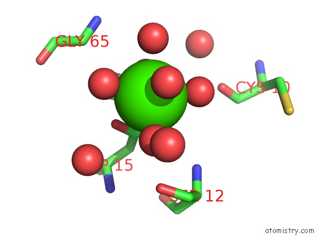
Mono view
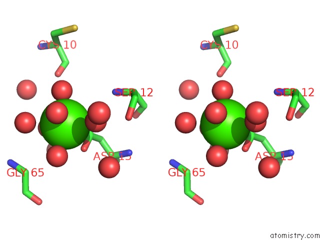
Stereo pair view
Mono view
Stereo pair view
A full contact list of Calcium with other atoms in the Ca binding
site number 1 of Ribonuclease T1 V16T Mutant within 5.0Å range:
|
Calcium binding site 2 out of 2 in 1i2g
Go back to
Calcium binding site 2 out
of 2 in the Ribonuclease T1 V16T Mutant
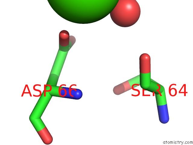
Mono view
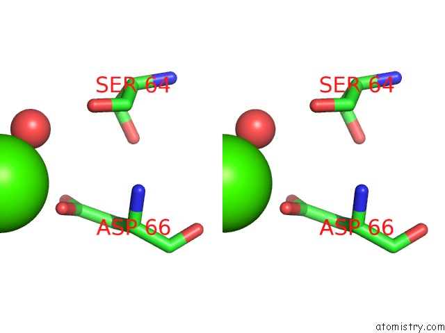
Stereo pair view
Mono view
Stereo pair view
A full contact list of Calcium with other atoms in the Ca binding
site number 2 of Ribonuclease T1 V16T Mutant within 5.0Å range:
|
Reference:
S.De Vos,
J.Backmann,
M.Prevost,
J.Steyaert,
R.Loris.
Hydrophobic Core Manipulations in Ribonuclease T1 Biochemistry V. 40 10140 2001.
ISSN: ISSN 0006-2960
PubMed: 11513591
DOI: 10.1021/BI010565N
Page generated: Mon Jul 7 15:45:55 2025
ISSN: ISSN 0006-2960
PubMed: 11513591
DOI: 10.1021/BI010565N
Last articles
Cl in 5H0ZCl in 5H0Y
Cl in 5GZK
Cl in 5GXZ
Cl in 5GWF
Cl in 5GXY
Cl in 5GXX
Cl in 5GWI
Cl in 5GWJ
Cl in 5GWB