Calcium in PDB 1i40: Structure of Inorganic Pyrophosphatase
Enzymatic activity of Structure of Inorganic Pyrophosphatase
All present enzymatic activity of Structure of Inorganic Pyrophosphatase:
3.6.1.1;
Protein crystallography data
The structure of Structure of Inorganic Pyrophosphatase, PDB code: 1i40
was solved by
V.R.Samygina,
A.N.Popov,
V.S.Lamzin,
S.M.Avaeva,
with X-Ray Crystallography technique. A brief refinement statistics is given in the table below:
Resolution Low / High (Å)
|
12.00 /
1.10
|
Space group
|
H 3 2
|
Cell size a, b, c (Å), α, β, γ (°)
|
109.520,
109.520,
75.080,
90.00,
90.00,
120.00
|
R / Rfree (%)
|
11.7 /
15.3
|
Other elements in 1i40:
The structure of Structure of Inorganic Pyrophosphatase also contains other interesting chemical elements:
Calcium Binding Sites:
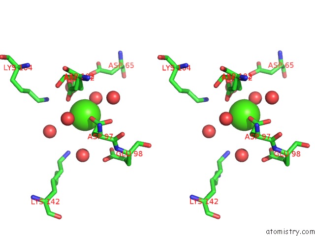
The binding sites of Calcium atom in the Structure of Inorganic Pyrophosphatase
(pdb code 1i40). This binding sites where shown within
5.0 Angstroms radius around Calcium atom.
In total 5 binding sites of Calcium where determined in the
Structure of Inorganic Pyrophosphatase, PDB code: 1i40:
Jump to Calcium binding site number:
1;
2;
3;
4;
5;
Calcium binding site 1 out
of 5 in 1i40
Go back to
Calcium Binding Sites List in 1i40
Calcium binding site 1 out
of 5 in the Structure of Inorganic Pyrophosphatase
Mono view
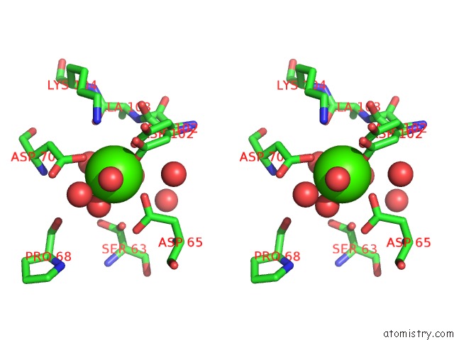
Stereo pair view
|
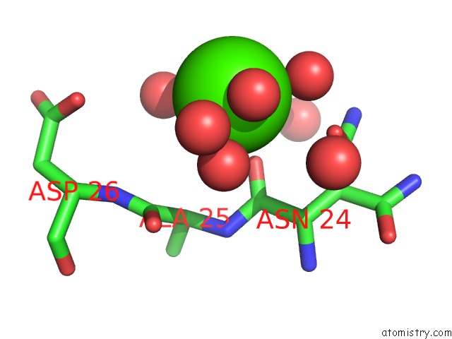
A full contact list of Calcium with other atoms in the Ca binding
site number 1 of Structure of Inorganic Pyrophosphatase within 5.0Å range:
probe
|
atom
|
residue
|
distance (Å)
|
B
|
Occ
|
A:Ca302
b:19.5
occ:0.70
|
OD1
|
A:ASP102
|
1.5
|
28.4
|
0.3
|
O
|
A:HOH310
|
2.1
|
25.1
|
1.0
|
O
|
A:HOH330
|
2.2
|
28.3
|
1.0
|
OD1
|
A:ASP102
|
2.3
|
20.9
|
0.7
|
OD1
|
A:ASP65
|
2.4
|
36.2
|
1.0
|
OD2
|
A:ASP70
|
2.4
|
23.8
|
1.0
|
O
|
A:HOH464
|
2.5
|
31.4
|
1.0
|
O
|
A:HOH323
|
2.6
|
18.2
|
1.0
|
CG
|
A:ASP102
|
2.6
|
26.0
|
0.3
|
OD2
|
A:ASP102
|
3.1
|
26.9
|
0.3
|
CG
|
A:ASP102
|
3.4
|
22.8
|
0.7
|
CG
|
A:ASP65
|
3.4
|
27.9
|
1.0
|
CG
|
A:ASP70
|
3.4
|
17.3
|
1.0
|
OD2
|
A:ASP65
|
3.7
|
25.6
|
1.0
|
OD2
|
A:ASP102
|
3.9
|
25.6
|
0.7
|
CB
|
A:ASP102
|
3.9
|
22.9
|
0.3
|
OG
|
A:SER63
|
4.0
|
21.6
|
0.3
|
O
|
A:ALA103
|
4.1
|
18.5
|
1.0
|
OD1
|
A:ASP70
|
4.1
|
20.1
|
1.0
|
O
|
A:HOH329
|
4.2
|
35.4
|
1.0
|
NZ
|
A:LYS104
|
4.2
|
22.6
|
1.0
|
O
|
A:HOH395
|
4.3
|
30.0
|
1.0
|
O
|
A:HOH383
|
4.3
|
37.3
|
0.8
|
CA
|
A:ASP102
|
4.4
|
21.7
|
0.3
|
CB
|
A:ASP70
|
4.4
|
14.5
|
1.0
|
C
|
A:ASP102
|
4.4
|
15.1
|
0.3
|
N
|
A:ALA103
|
4.5
|
16.7
|
1.0
|
O
|
A:PRO68
|
4.6
|
16.5
|
1.0
|
CB
|
A:ASP102
|
4.6
|
19.3
|
0.7
|
O
|
A:ASP102
|
4.7
|
20.2
|
0.3
|
N
|
A:ASP70
|
4.7
|
12.8
|
1.0
|
CB
|
A:ASP65
|
4.7
|
24.1
|
1.0
|
O
|
A:HOH382
|
4.8
|
20.6
|
0.5
|
CA
|
A:ASP102
|
4.9
|
20.0
|
0.7
|
|
Calcium binding site 2 out
of 5 in 1i40
Go back to
Calcium Binding Sites List in 1i40
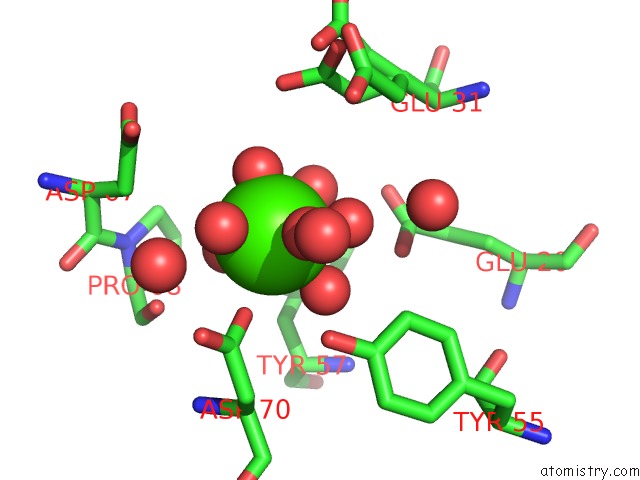
Calcium binding site 2 out
of 5 in the Structure of Inorganic Pyrophosphatase
Mono view
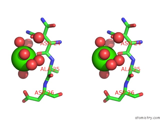
Stereo pair view
|
A full contact list of Calcium with other atoms in the Ca binding
site number 2 of Structure of Inorganic Pyrophosphatase within 5.0Å range:
probe
|
atom
|
residue
|
distance (Å)
|
B
|
Occ
|
A:Ca303
b:18.4
occ:0.50
|
O
|
A:HOH506
|
2.0
|
32.6
|
0.7
|
O
|
A:HOH389
|
2.3
|
33.2
|
0.5
|
O
|
A:HOH332
|
2.5
|
21.3
|
0.5
|
O
|
A:ASN24
|
2.6
|
21.4
|
1.0
|
O
|
A:HOH507
|
2.6
|
15.0
|
0.3
|
O
|
A:HOH470
|
2.9
|
50.6
|
1.0
|
O
|
A:HOH359
|
3.2
|
41.4
|
0.5
|
OD1
|
A:ASN24
|
3.3
|
35.1
|
0.6
|
C
|
A:ASN24
|
3.5
|
16.0
|
1.0
|
CG
|
A:ASN24
|
3.8
|
26.9
|
0.6
|
ND2
|
A:ASN24
|
4.2
|
35.5
|
0.6
|
O
|
A:HOH572
|
4.2
|
44.0
|
1.0
|
N
|
A:ALA25
|
4.2
|
13.2
|
1.0
|
CA
|
A:ALA25
|
4.3
|
12.8
|
1.0
|
C
|
A:ALA25
|
4.3
|
12.2
|
1.0
|
CA
|
A:ASN24
|
4.4
|
21.0
|
1.0
|
O
|
A:HOH554
|
4.5
|
24.0
|
0.3
|
O
|
A:ALA25
|
4.5
|
16.1
|
1.0
|
CB
|
A:ASN24
|
4.6
|
27.9
|
0.6
|
CB
|
A:ASN24
|
4.6
|
26.4
|
0.4
|
OD1
|
A:ASP26
|
4.7
|
24.0
|
1.0
|
N
|
A:ASP26
|
4.8
|
12.8
|
1.0
|
|
Calcium binding site 3 out
of 5 in 1i40
Go back to
Calcium Binding Sites List in 1i40
Calcium binding site 3 out
of 5 in the Structure of Inorganic Pyrophosphatase
Mono view
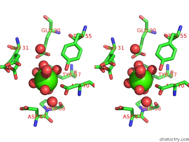
Stereo pair view
|
A full contact list of Calcium with other atoms in the Ca binding
site number 3 of Structure of Inorganic Pyrophosphatase within 5.0Å range:
probe
|
atom
|
residue
|
distance (Å)
|
B
|
Occ
|
A:Ca304
b:22.7
occ:0.60
|
O
|
A:HOH377
|
1.9
|
42.4
|
1.0
|
O
|
A:HOH322
|
2.1
|
22.9
|
1.0
|
O
|
A:HOH329
|
2.2
|
35.4
|
1.0
|
O
|
A:HOH509
|
2.3
|
18.0
|
0.4
|
O
|
A:HOH315
|
2.4
|
20.2
|
0.6
|
O
|
A:HOH508
|
2.5
|
28.3
|
0.6
|
OD1
|
A:ASP70
|
2.6
|
20.1
|
1.0
|
O
|
A:HOH539
|
2.7
|
27.1
|
0.6
|
CG
|
A:ASP70
|
3.8
|
17.3
|
1.0
|
O
|
A:HOH394
|
4.1
|
16.2
|
0.5
|
OD2
|
A:ASP70
|
4.3
|
23.8
|
1.0
|
OE1
|
A:GLU20
|
4.4
|
15.3
|
1.0
|
O
|
A:HOH310
|
4.4
|
25.1
|
1.0
|
O
|
A:PRO68
|
4.5
|
16.5
|
1.0
|
OH
|
A:TYR55
|
4.5
|
18.0
|
1.0
|
OE2
|
A:GLU31
|
4.6
|
43.8
|
0.5
|
CB
|
A:ASP67
|
4.7
|
25.5
|
1.0
|
O
|
A:HOH314
|
4.8
|
33.9
|
1.0
|
CB
|
A:TYR57
|
4.8
|
11.8
|
1.0
|
CB
|
A:ASP70
|
4.9
|
14.5
|
1.0
|
OE2
|
A:GLU20
|
4.9
|
13.9
|
1.0
|
CE2
|
A:TYR55
|
5.0
|
15.0
|
1.0
|
|
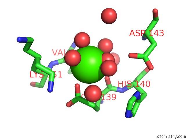
Calcium binding site 4 out
of 5 in 1i40
Go back to
Calcium Binding Sites List in 1i40
Calcium binding site 4 out
of 5 in the Structure of Inorganic Pyrophosphatase
Mono view
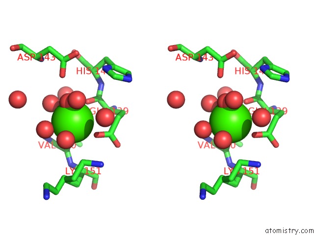
Stereo pair view
|
A full contact list of Calcium with other atoms in the Ca binding
site number 4 of Structure of Inorganic Pyrophosphatase within 5.0Å range:
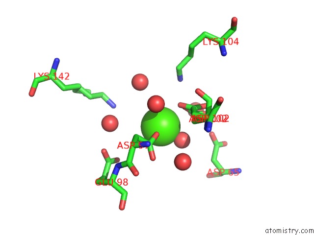
probe
|
atom
|
residue
|
distance (Å)
|
B
|
Occ
|
A:Ca305
b:20.2
occ:0.70
|
O
|
A:HOH452
|
1.8
|
21.7
|
0.6
|
O
|
A:VAL150
|
2.3
|
21.6
|
1.0
|
OE2
|
A:GLU139
|
2.3
|
30.3
|
1.0
|
O
|
A:HOH503
|
2.4
|
24.4
|
0.5
|
O
|
A:HOH531
|
2.4
|
31.2
|
0.6
|
O
|
A:GLU139
|
2.5
|
15.7
|
1.0
|
O
|
A:HOH347
|
2.5
|
22.7
|
0.8
|
O
|
A:HOH396
|
2.7
|
20.7
|
1.0
|
CD
|
A:GLU139
|
3.3
|
19.3
|
1.0
|
C
|
A:VAL150
|
3.6
|
19.4
|
1.0
|
C
|
A:GLU139
|
3.6
|
13.5
|
1.0
|
CE
|
A:LYS151
|
3.8
|
53.5
|
0.7
|
CG
|
A:GLU139
|
3.9
|
18.7
|
1.0
|
OD1
|
A:ASP143
|
4.0
|
15.6
|
1.0
|
CA
|
A:GLU139
|
4.1
|
13.9
|
1.0
|
N
|
A:VAL150
|
4.2
|
20.3
|
1.0
|
O
|
A:HOH493
|
4.2
|
37.2
|
0.6
|
OE1
|
A:GLU139
|
4.3
|
21.4
|
1.0
|
O
|
A:HOH335
|
4.4
|
15.4
|
1.0
|
CA
|
A:VAL150
|
4.4
|
19.4
|
1.0
|
N
|
A:LYS151
|
4.5
|
20.4
|
1.0
|
CA
|
A:LYS151
|
4.6
|
20.8
|
1.0
|
CB
|
A:GLU139
|
4.6
|
15.2
|
1.0
|
NZ
|
A:LYS151
|
4.6
|
29.5
|
0.7
|
N
|
A:HIS140
|
4.8
|
12.8
|
1.0
|
CG
|
A:LYS151
|
4.8
|
32.9
|
0.3
|
CB
|
A:VAL150
|
4.8
|
19.9
|
1.0
|
CG
|
A:LYS151
|
4.9
|
36.4
|
0.7
|
CD
|
A:LYS151
|
4.9
|
51.3
|
0.7
|
O
|
A:HOH342
|
5.0
|
16.2
|
1.0
|
|
Calcium binding site 5 out
of 5 in 1i40
Go back to
Calcium Binding Sites List in 1i40
Calcium binding site 5 out
of 5 in the Structure of Inorganic Pyrophosphatase
Mono view
Stereo pair view
|
A full contact list of Calcium with other atoms in the Ca binding
site number 5 of Structure of Inorganic Pyrophosphatase within 5.0Å range:
probe
|
atom
|
residue
|
distance (Å)
|
B
|
Occ
|
A:Ca306
b:23.3
occ:0.20
|
OD2
|
A:ASP97
|
2.2
|
35.6
|
1.0
|
O
|
A:HOH383
|
2.3
|
37.3
|
0.8
|
OD2
|
A:ASP102
|
2.8
|
26.9
|
0.3
|
OD2
|
A:ASP102
|
2.9
|
25.6
|
0.7
|
CG
|
A:ASP97
|
3.3
|
23.9
|
1.0
|
O
|
A:HOH482
|
3.4
|
25.5
|
0.7
|
CG
|
A:ASP102
|
3.5
|
26.0
|
0.3
|
CB
|
A:ASP102
|
3.7
|
22.9
|
0.3
|
CG
|
A:ASP102
|
3.8
|
22.8
|
0.7
|
OD1
|
A:ASP97
|
4.0
|
25.3
|
1.0
|
O
|
A:HOH310
|
4.2
|
25.1
|
1.0
|
O
|
A:HOH448
|
4.2
|
32.9
|
1.0
|
CB
|
A:ASP97
|
4.3
|
23.5
|
1.0
|
OD1
|
A:ASP102
|
4.4
|
28.4
|
0.3
|
CB
|
A:ASP102
|
4.5
|
19.3
|
0.7
|
O
|
A:HOH368
|
4.5
|
24.6
|
0.9
|
OD1
|
A:ASP102
|
4.5
|
20.9
|
0.7
|
O
|
A:HOH481
|
4.6
|
42.9
|
1.0
|
NZ
|
A:LYS104
|
4.7
|
22.6
|
1.0
|
NZ
|
A:LYS142
|
4.8
|
27.1
|
0.7
|
OD1
|
A:ASP65
|
4.8
|
36.2
|
1.0
|
OE2
|
A:GLU98
|
5.0
|
34.4
|
1.0
|
|
Reference:
V.R.Samygina,
A.N.Popov,
E.V.Rodina,
N.N.Vorobyeva,
V.S.Lamzin,
K.M.Polyakov,
S.A.Kurilova,
T.I.Nazarova,
S.M.Avaeva.
The Structures of Escherichia Coli Inorganic Pyrophosphatase Complexed with Ca(2+) or Capp(I) at Atomic Resolution and Their Mechanistic Implications. J.Mol.Biol. V. 314 633 2001.
ISSN: ISSN 0022-2836
PubMed: 11846572
DOI: 10.1006/JMBI.2001.5149
Page generated: Thu Jul 11 10:15:29 2024
|