Calcium »
PDB 1i73-1iqi »
1i7o »
Calcium in PDB 1i7o: Crystal Structure of Hpce
Enzymatic activity of Crystal Structure of Hpce
Protein crystallography data
The structure of Crystal Structure of Hpce, PDB code: 1i7o
was solved by
J.R.H.Tame,
K.Namba,
E.J.Dodson,
D.I.Roper,
with X-Ray Crystallography technique. A brief refinement statistics is given in the table below:
| Resolution Low / High (Å) | 15.00 / 1.70 |
| Space group | P 21 21 21 |
| Cell size a, b, c (Å), α, β, γ (°) | 126.084, 138.201, 103.973, 90.00, 90.00, 90.00 |
| R / Rfree (%) | 25.1 / 27.8 |
Calcium Binding Sites:
The binding sites of Calcium atom in the Crystal Structure of Hpce
(pdb code 1i7o). This binding sites where shown within
5.0 Angstroms radius around Calcium atom.
In total 4 binding sites of Calcium where determined in the Crystal Structure of Hpce, PDB code: 1i7o:
Jump to Calcium binding site number: 1; 2; 3; 4;
In total 4 binding sites of Calcium where determined in the Crystal Structure of Hpce, PDB code: 1i7o:
Jump to Calcium binding site number: 1; 2; 3; 4;
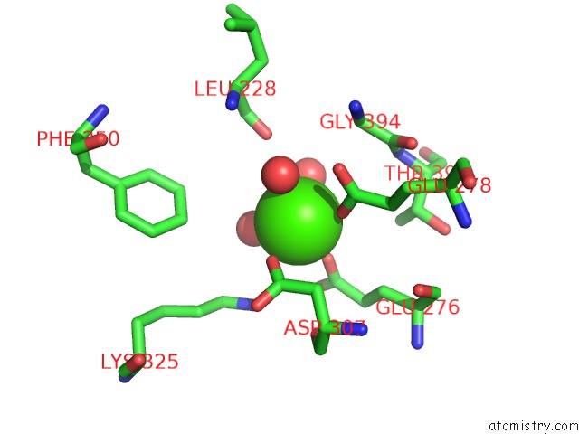
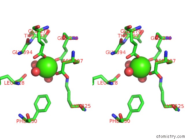
Calcium binding site 1 out of 4 in 1i7o
Go back to
Calcium binding site 1 out
of 4 in the Crystal Structure of Hpce
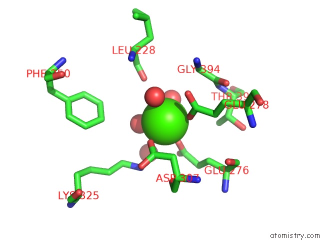
Mono view
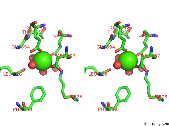
Stereo pair view
Mono view
Stereo pair view
A full contact list of Calcium with other atoms in the Ca binding
site number 1 of Crystal Structure of Hpce within 5.0Å range:
|
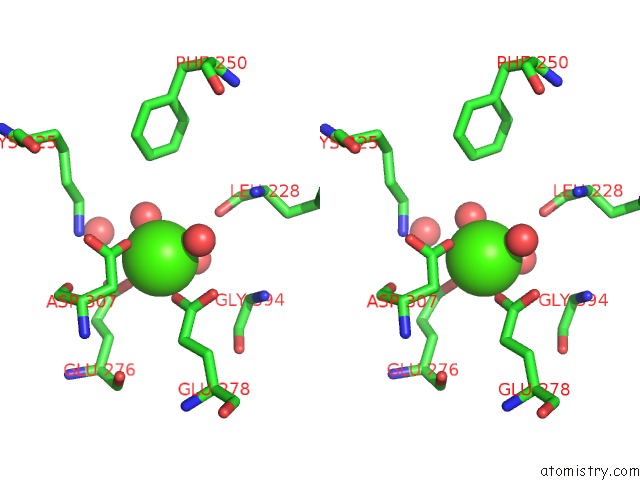
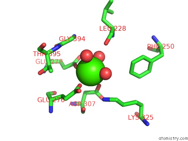
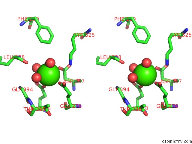
Calcium binding site 2 out of 4 in 1i7o
Go back to
Calcium binding site 2 out
of 4 in the Crystal Structure of Hpce
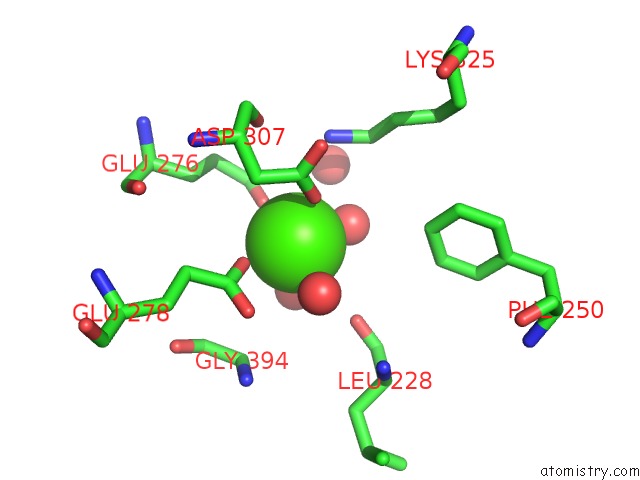
Mono view
Stereo pair view
Mono view
Stereo pair view
A full contact list of Calcium with other atoms in the Ca binding
site number 2 of Crystal Structure of Hpce within 5.0Å range:
|
Calcium binding site 3 out of 4 in 1i7o
Go back to
Calcium binding site 3 out
of 4 in the Crystal Structure of Hpce
Mono view
Stereo pair view
Mono view
Stereo pair view
A full contact list of Calcium with other atoms in the Ca binding
site number 3 of Crystal Structure of Hpce within 5.0Å range:
|
Calcium binding site 4 out of 4 in 1i7o
Go back to
Calcium binding site 4 out
of 4 in the Crystal Structure of Hpce
Mono view
Stereo pair view
Mono view
Stereo pair view
A full contact list of Calcium with other atoms in the Ca binding
site number 4 of Crystal Structure of Hpce within 5.0Å range:
|
Reference:
J.R.H.Tame,
K.Namba,
E.J.Dodson,
D.I.Roper.
The Crystal Structure of Hpce, A Multi-Functional Enzyme Fold To Be Published.
Page generated: Mon Jul 7 15:47:14 2025
Last articles
Cl in 5KTPCl in 5KTN
Cl in 5KT9
Cl in 5KT8
Cl in 5KTM
Cl in 5KSZ
Cl in 5KT1
Cl in 5KSR
Cl in 5KSW
Cl in 5KSS