Calcium »
PDB 1i73-1iqi »
1i9b »
Calcium in PDB 1i9b: X-Ray Structure of Acetylcholine Binding Protein (Achbp)
Protein crystallography data
The structure of X-Ray Structure of Acetylcholine Binding Protein (Achbp), PDB code: 1i9b
was solved by
K.Brejc,
W.J.Van Dijk,
R.Klaassen,
M.Schuurmans,
J.Van Der Oost,
A.B.Smit,
T.K.Sixma,
with X-Ray Crystallography technique. A brief refinement statistics is given in the table below:
| Resolution Low / High (Å) | 19.97 / 2.70 |
| Space group | P 42 21 2 |
| Cell size a, b, c (Å), α, β, γ (°) | 141.660, 141.660, 120.870, 90.00, 90.00, 90.00 |
| R / Rfree (%) | 26 / 29.7 |
Calcium Binding Sites:
The binding sites of Calcium atom in the X-Ray Structure of Acetylcholine Binding Protein (Achbp)
(pdb code 1i9b). This binding sites where shown within
5.0 Angstroms radius around Calcium atom.
In total 10 binding sites of Calcium where determined in the X-Ray Structure of Acetylcholine Binding Protein (Achbp), PDB code: 1i9b:
Jump to Calcium binding site number: 1; 2; 3; 4; 5; 6; 7; 8; 9; 10;
In total 10 binding sites of Calcium where determined in the X-Ray Structure of Acetylcholine Binding Protein (Achbp), PDB code: 1i9b:
Jump to Calcium binding site number: 1; 2; 3; 4; 5; 6; 7; 8; 9; 10;
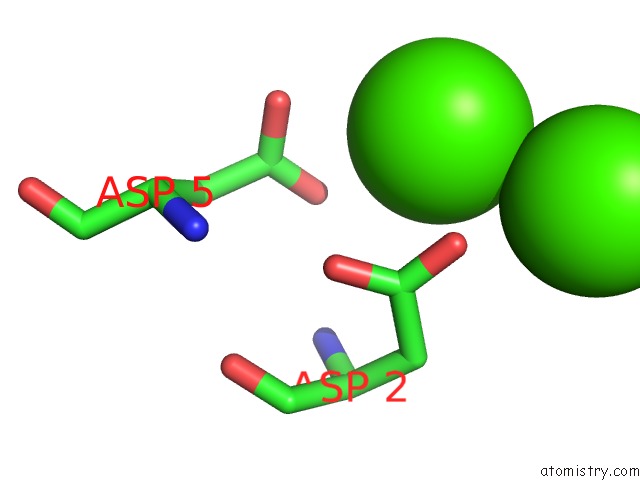
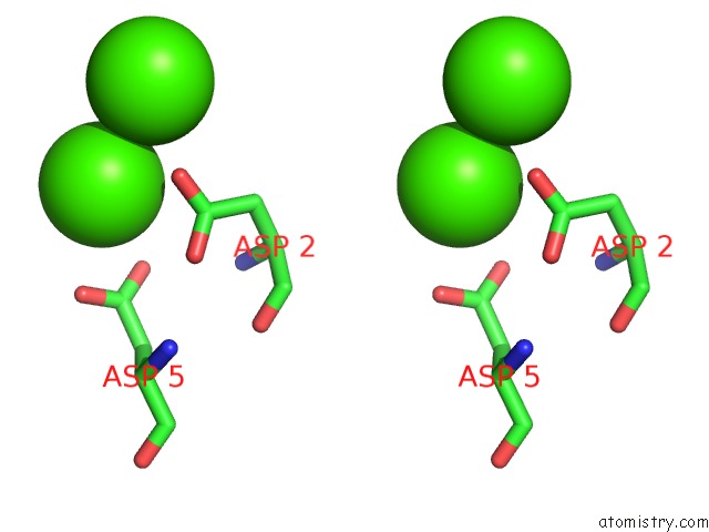
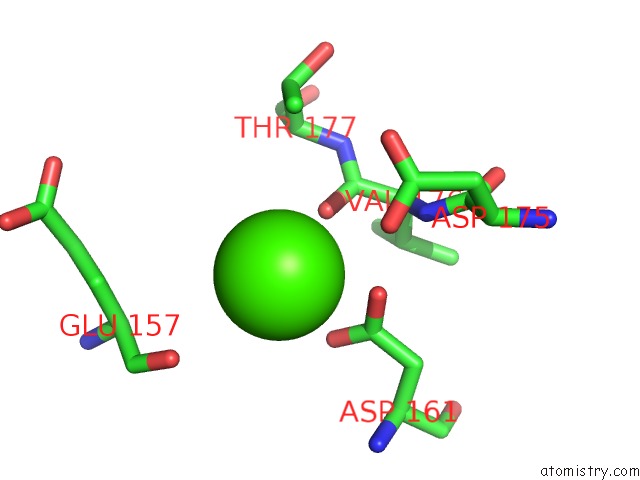
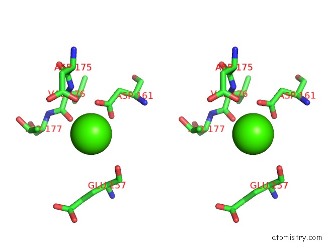
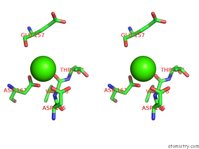
Calcium binding site 1 out of 10 in 1i9b
Go back to
Calcium binding site 1 out
of 10 in the X-Ray Structure of Acetylcholine Binding Protein (Achbp)
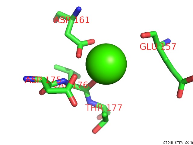
Mono view
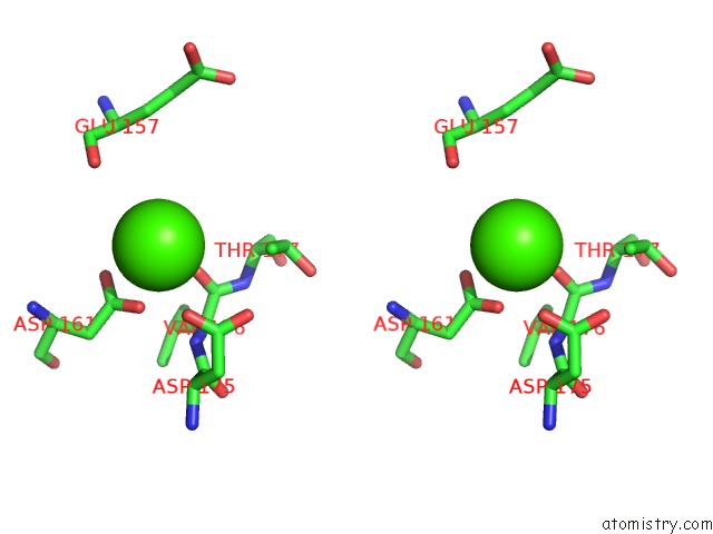
Stereo pair view
Mono view
Stereo pair view
A full contact list of Calcium with other atoms in the Ca binding
site number 1 of X-Ray Structure of Acetylcholine Binding Protein (Achbp) within 5.0Å range:
|
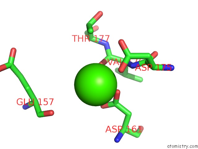
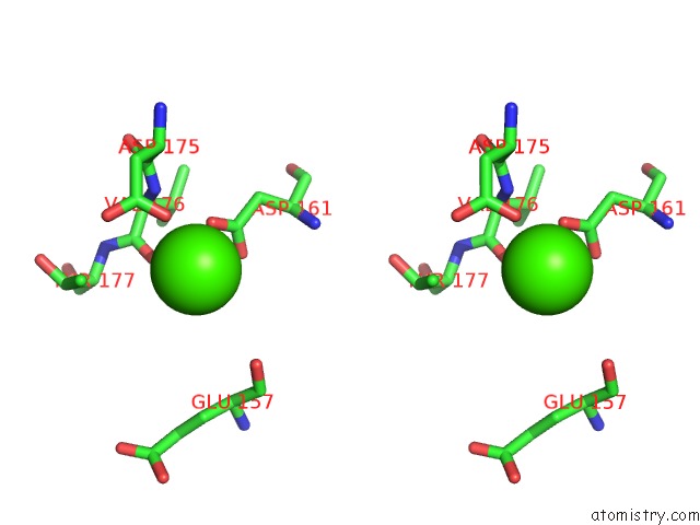
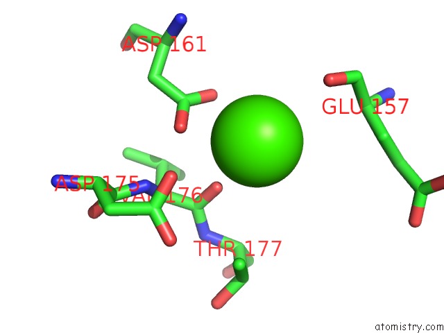
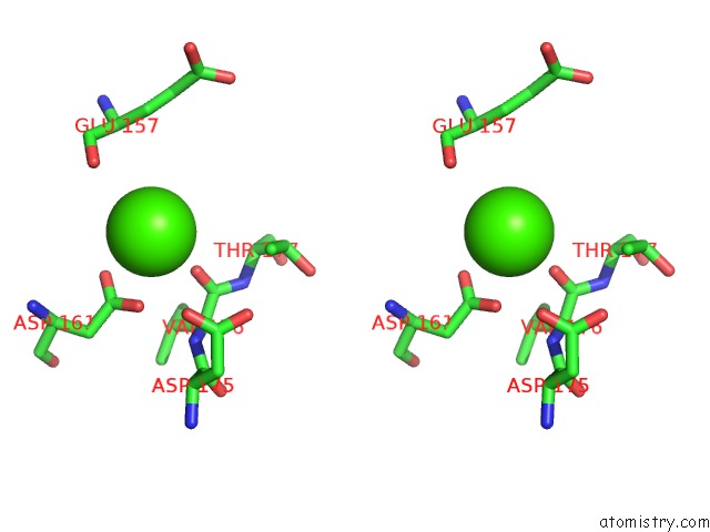
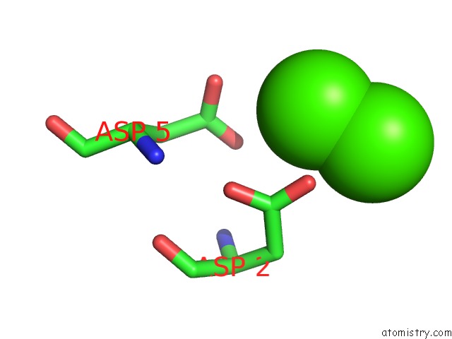
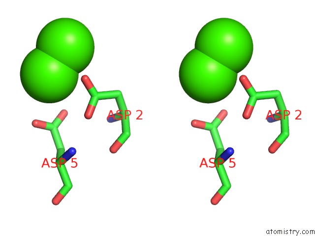
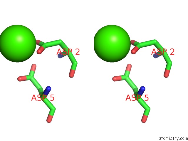
Calcium binding site 2 out of 10 in 1i9b
Go back to
Calcium binding site 2 out
of 10 in the X-Ray Structure of Acetylcholine Binding Protein (Achbp)
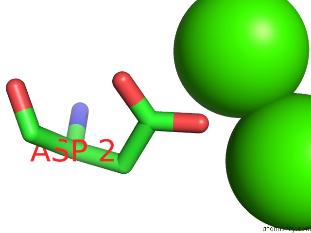
Mono view
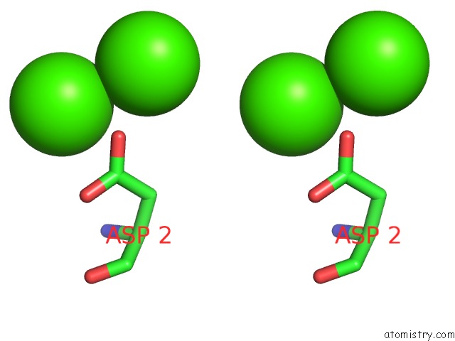
Stereo pair view
Mono view
Stereo pair view
A full contact list of Calcium with other atoms in the Ca binding
site number 2 of X-Ray Structure of Acetylcholine Binding Protein (Achbp) within 5.0Å range:
|
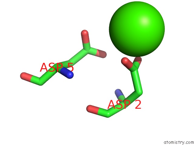
Calcium binding site 3 out of 10 in 1i9b
Go back to
Calcium binding site 3 out
of 10 in the X-Ray Structure of Acetylcholine Binding Protein (Achbp)
Mono view
Stereo pair view
Mono view
Stereo pair view
A full contact list of Calcium with other atoms in the Ca binding
site number 3 of X-Ray Structure of Acetylcholine Binding Protein (Achbp) within 5.0Å range:
|
Calcium binding site 4 out of 10 in 1i9b
Go back to
Calcium binding site 4 out
of 10 in the X-Ray Structure of Acetylcholine Binding Protein (Achbp)
Mono view
Stereo pair view
Mono view
Stereo pair view
A full contact list of Calcium with other atoms in the Ca binding
site number 4 of X-Ray Structure of Acetylcholine Binding Protein (Achbp) within 5.0Å range:
|
Calcium binding site 5 out of 10 in 1i9b
Go back to
Calcium binding site 5 out
of 10 in the X-Ray Structure of Acetylcholine Binding Protein (Achbp)
Mono view
Stereo pair view
Mono view
Stereo pair view
A full contact list of Calcium with other atoms in the Ca binding
site number 5 of X-Ray Structure of Acetylcholine Binding Protein (Achbp) within 5.0Å range:
|
Calcium binding site 6 out of 10 in 1i9b
Go back to
Calcium binding site 6 out
of 10 in the X-Ray Structure of Acetylcholine Binding Protein (Achbp)
Mono view
Stereo pair view
Mono view
Stereo pair view
A full contact list of Calcium with other atoms in the Ca binding
site number 6 of X-Ray Structure of Acetylcholine Binding Protein (Achbp) within 5.0Å range:
|
Calcium binding site 7 out of 10 in 1i9b
Go back to
Calcium binding site 7 out
of 10 in the X-Ray Structure of Acetylcholine Binding Protein (Achbp)

Mono view
Stereo pair view
Mono view
Stereo pair view
A full contact list of Calcium with other atoms in the Ca binding
site number 7 of X-Ray Structure of Acetylcholine Binding Protein (Achbp) within 5.0Å range:
|
Calcium binding site 8 out of 10 in 1i9b
Go back to
Calcium binding site 8 out
of 10 in the X-Ray Structure of Acetylcholine Binding Protein (Achbp)
Mono view
Stereo pair view
Mono view
Stereo pair view
A full contact list of Calcium with other atoms in the Ca binding
site number 8 of X-Ray Structure of Acetylcholine Binding Protein (Achbp) within 5.0Å range:
|
Calcium binding site 9 out of 10 in 1i9b
Go back to
Calcium binding site 9 out
of 10 in the X-Ray Structure of Acetylcholine Binding Protein (Achbp)

Mono view

Stereo pair view
Mono view
Stereo pair view
A full contact list of Calcium with other atoms in the Ca binding
site number 9 of X-Ray Structure of Acetylcholine Binding Protein (Achbp) within 5.0Å range:
|
Calcium binding site 10 out of 10 in 1i9b
Go back to
Calcium binding site 10 out
of 10 in the X-Ray Structure of Acetylcholine Binding Protein (Achbp)
Mono view
Stereo pair view
Mono view
Stereo pair view
A full contact list of Calcium with other atoms in the Ca binding
site number 10 of X-Ray Structure of Acetylcholine Binding Protein (Achbp) within 5.0Å range:
|
Reference:
K.Brejc,
W.J.Van Dijk,
R.V.Klaassen,
M.Schuurmans,
J.Van Der Oost,
A.B.Smit,
T.K.Sixma.
Crystal Structure of An Ach-Binding Protein Reveals the Ligand-Binding Domain of Nicotinic Receptors. Nature V. 411 269 2001.
ISSN: ISSN 0028-0836
PubMed: 11357122
DOI: 10.1038/35077011
Page generated: Mon Jul 7 15:48:29 2025
ISSN: ISSN 0028-0836
PubMed: 11357122
DOI: 10.1038/35077011
Last articles
Cl in 5R9SCl in 5R9U
Cl in 5R9T
Cl in 5R9R
Cl in 5R9P
Cl in 5R9Q
Cl in 5R9O
Cl in 5R9N
Cl in 5R9M
Cl in 5R9K