Calcium »
PDB 1i73-1iqi »
1id5 »
Calcium in PDB 1id5: Crystal Structure of Bovine Thrombin Complex with Protease Inhibitor Ecotin
Enzymatic activity of Crystal Structure of Bovine Thrombin Complex with Protease Inhibitor Ecotin
All present enzymatic activity of Crystal Structure of Bovine Thrombin Complex with Protease Inhibitor Ecotin:
3.4.21.5;
3.4.21.5;
Protein crystallography data
The structure of Crystal Structure of Bovine Thrombin Complex with Protease Inhibitor Ecotin, PDB code: 1id5
was solved by
S.X.Wang,
R.J.Fletterick,
with X-Ray Crystallography technique. A brief refinement statistics is given in the table below:
| Resolution Low / High (Å) | 6.00 / 2.50 |
| Space group | C 2 2 21 |
| Cell size a, b, c (Å), α, β, γ (°) | 88.517, 165.377, 83.326, 90.00, 90.00, 90.00 |
| R / Rfree (%) | 20.3 / 26.4 |
Calcium Binding Sites:
The binding sites of Calcium atom in the Crystal Structure of Bovine Thrombin Complex with Protease Inhibitor Ecotin
(pdb code 1id5). This binding sites where shown within
5.0 Angstroms radius around Calcium atom.
In total only one binding site of Calcium was determined in the Crystal Structure of Bovine Thrombin Complex with Protease Inhibitor Ecotin, PDB code: 1id5:
In total only one binding site of Calcium was determined in the Crystal Structure of Bovine Thrombin Complex with Protease Inhibitor Ecotin, PDB code: 1id5:
Calcium binding site 1 out of 1 in 1id5
Go back to
Calcium binding site 1 out
of 1 in the Crystal Structure of Bovine Thrombin Complex with Protease Inhibitor Ecotin
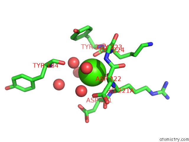
Mono view
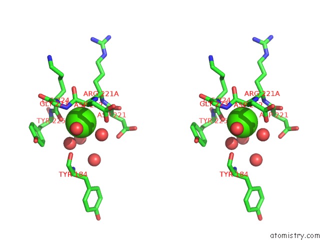
Stereo pair view
Mono view
Stereo pair view
A full contact list of Calcium with other atoms in the Ca binding
site number 1 of Crystal Structure of Bovine Thrombin Complex with Protease Inhibitor Ecotin within 5.0Å range:
|
Reference:
S.X.Wang,
C.T.Esmon,
R.J.Fletterick.
Crystal Structure of Thrombin-Ecotin Reveals Conformational Changes and Extended Interactions. Biochemistry V. 40 10038 2001.
ISSN: ISSN 0006-2960
PubMed: 11513582
DOI: 10.1021/BI010712H
Page generated: Thu Jul 11 10:19:35 2024
ISSN: ISSN 0006-2960
PubMed: 11513582
DOI: 10.1021/BI010712H
Last articles
Zn in 9J0NZn in 9J0O
Zn in 9J0P
Zn in 9FJX
Zn in 9EKB
Zn in 9C0F
Zn in 9CAH
Zn in 9CH0
Zn in 9CH3
Zn in 9CH1