Calcium »
PDB 1iqj-1j1n »
1iyi »
Calcium in PDB 1iyi: Crystal Structure of Hematopoietic Prostaglandin D Synthase
Enzymatic activity of Crystal Structure of Hematopoietic Prostaglandin D Synthase
All present enzymatic activity of Crystal Structure of Hematopoietic Prostaglandin D Synthase:
5.3.99.2;
5.3.99.2;
Protein crystallography data
The structure of Crystal Structure of Hematopoietic Prostaglandin D Synthase, PDB code: 1iyi
was solved by
T.Inoue,
with X-Ray Crystallography technique. A brief refinement statistics is given in the table below:
| Resolution Low / High (Å) | 37.29 / 1.80 |
| Space group | P 1 21 1 |
| Cell size a, b, c (Å), α, β, γ (°) | 48.784, 47.263, 183.810, 90.00, 97.83, 90.00 |
| R / Rfree (%) | 18.8 / 22 |
Calcium Binding Sites:
The binding sites of Calcium atom in the Crystal Structure of Hematopoietic Prostaglandin D Synthase
(pdb code 1iyi). This binding sites where shown within
5.0 Angstroms radius around Calcium atom.
In total 2 binding sites of Calcium where determined in the Crystal Structure of Hematopoietic Prostaglandin D Synthase, PDB code: 1iyi:
Jump to Calcium binding site number: 1; 2;
In total 2 binding sites of Calcium where determined in the Crystal Structure of Hematopoietic Prostaglandin D Synthase, PDB code: 1iyi:
Jump to Calcium binding site number: 1; 2;
Calcium binding site 1 out of 2 in 1iyi
Go back to
Calcium binding site 1 out
of 2 in the Crystal Structure of Hematopoietic Prostaglandin D Synthase
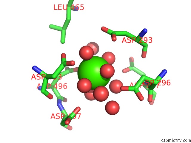
Mono view
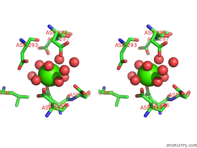
Stereo pair view
Mono view
Stereo pair view
A full contact list of Calcium with other atoms in the Ca binding
site number 1 of Crystal Structure of Hematopoietic Prostaglandin D Synthase within 5.0Å range:
|
Calcium binding site 2 out of 2 in 1iyi
Go back to
Calcium binding site 2 out
of 2 in the Crystal Structure of Hematopoietic Prostaglandin D Synthase
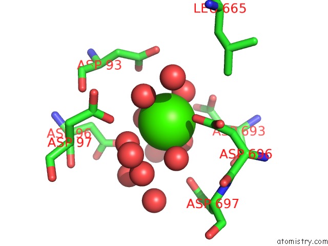
Mono view
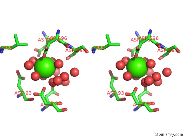
Stereo pair view
Mono view
Stereo pair view
A full contact list of Calcium with other atoms in the Ca binding
site number 2 of Crystal Structure of Hematopoietic Prostaglandin D Synthase within 5.0Å range:
|
Reference:
T.Inoue,
D.Irikura,
N.Okazaki,
S.Kinugasa,
H.Matsumura,
N.Uodome,
M.Yamamoto,
T.Kumasaka,
M.Miyano,
Y.Kai,
Y.Urade.
Mechanism of Metal Activation of Human Hematopoietic Prostaglandin D Synthase Nat.Struct.Biol. V. 10 291 2003.
ISSN: ISSN 1072-8368
PubMed: 12627223
DOI: 10.1038/NSB907
Page generated: Thu Jul 11 10:28:18 2024
ISSN: ISSN 1072-8368
PubMed: 12627223
DOI: 10.1038/NSB907
Last articles
Zn in 9MJ5Zn in 9HNW
Zn in 9G0L
Zn in 9FNE
Zn in 9DZN
Zn in 9E0I
Zn in 9D32
Zn in 9DAK
Zn in 8ZXC
Zn in 8ZUF