Calcium »
PDB 1iqj-1j1n »
1j18 »
Calcium in PDB 1j18: Crystal Structure of A Beta-Amylase From Bacillus Cereus Var. Mycoides Cocrystallized with Maltose
Enzymatic activity of Crystal Structure of A Beta-Amylase From Bacillus Cereus Var. Mycoides Cocrystallized with Maltose
All present enzymatic activity of Crystal Structure of A Beta-Amylase From Bacillus Cereus Var. Mycoides Cocrystallized with Maltose:
3.2.1.2;
3.2.1.2;
Protein crystallography data
The structure of Crystal Structure of A Beta-Amylase From Bacillus Cereus Var. Mycoides Cocrystallized with Maltose, PDB code: 1j18
was solved by
H.Miyake,
G.Kurisu,
M.Kusunoki,
S.Nishimura,
S.Kitamura,
Y.Nitta,
with X-Ray Crystallography technique. A brief refinement statistics is given in the table below:
| Resolution Low / High (Å) | 29.00 / 2.00 |
| Space group | P 1 21 1 |
| Cell size a, b, c (Å), α, β, γ (°) | 56.808, 89.331, 65.394, 90.00, 102.32, 90.00 |
| R / Rfree (%) | 18.1 / 22.1 |
Calcium Binding Sites:
The binding sites of Calcium atom in the Crystal Structure of A Beta-Amylase From Bacillus Cereus Var. Mycoides Cocrystallized with Maltose
(pdb code 1j18). This binding sites where shown within
5.0 Angstroms radius around Calcium atom.
In total only one binding site of Calcium was determined in the Crystal Structure of A Beta-Amylase From Bacillus Cereus Var. Mycoides Cocrystallized with Maltose, PDB code: 1j18:
In total only one binding site of Calcium was determined in the Crystal Structure of A Beta-Amylase From Bacillus Cereus Var. Mycoides Cocrystallized with Maltose, PDB code: 1j18:
Calcium binding site 1 out of 1 in 1j18
Go back to
Calcium binding site 1 out
of 1 in the Crystal Structure of A Beta-Amylase From Bacillus Cereus Var. Mycoides Cocrystallized with Maltose
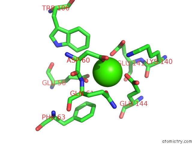
Mono view
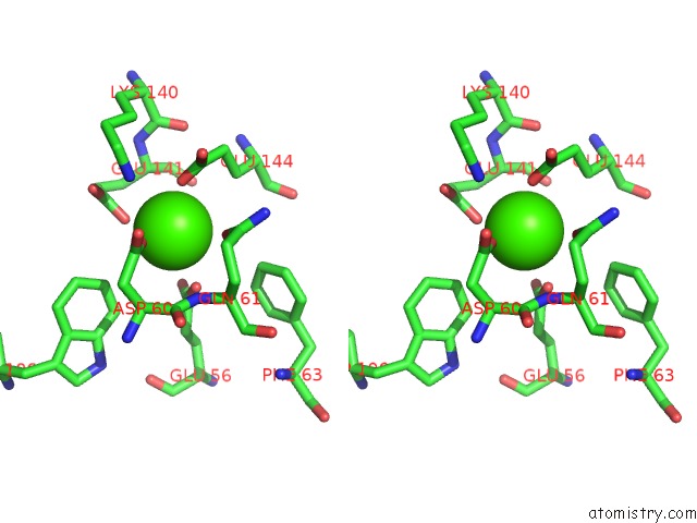
Stereo pair view
Mono view
Stereo pair view
A full contact list of Calcium with other atoms in the Ca binding
site number 1 of Crystal Structure of A Beta-Amylase From Bacillus Cereus Var. Mycoides Cocrystallized with Maltose within 5.0Å range:
|
Reference:
H.Miyake,
G.Kurisu,
M.Kusunoki,
S.Nishimura,
S.Kitamura,
Y.Nitta.
Crystal Structure of A Catalytic Site Mutant of Beta-Amylase From Bacillus Cereus Var. Mycoides Cocrystallized with Maltopentaose Biochemistry V. 42 5574 2003.
ISSN: ISSN 0006-2960
PubMed: 12741813
DOI: 10.1021/BI020712X
Page generated: Thu Jul 11 10:33:28 2024
ISSN: ISSN 0006-2960
PubMed: 12741813
DOI: 10.1021/BI020712X
Last articles
Zn in 9MJ5Zn in 9HNW
Zn in 9G0L
Zn in 9FNE
Zn in 9DZN
Zn in 9E0I
Zn in 9D32
Zn in 9DAK
Zn in 8ZXC
Zn in 8ZUF