Calcium »
PDB 1iqj-1j1n »
1j1e »
Calcium in PDB 1j1e: Crystal Structure of the 52KDA Domain of Human Cardiac Troponin in the CA2+ Saturated Form
Protein crystallography data
The structure of Crystal Structure of the 52KDA Domain of Human Cardiac Troponin in the CA2+ Saturated Form, PDB code: 1j1e
was solved by
S.Takeda,
A.Yamashita,
K.Maeda,
Y.Maeda,
with X-Ray Crystallography technique. A brief refinement statistics is given in the table below:
| Resolution Low / High (Å) | 20.00 / 3.30 |
| Space group | P 1 21 1 |
| Cell size a, b, c (Å), α, β, γ (°) | 48.299, 169.506, 68.538, 90.00, 102.38, 90.00 |
| R / Rfree (%) | 25.1 / 30.8 |
Calcium Binding Sites:
The binding sites of Calcium atom in the Crystal Structure of the 52KDA Domain of Human Cardiac Troponin in the CA2+ Saturated Form
(pdb code 1j1e). This binding sites where shown within
5.0 Angstroms radius around Calcium atom.
In total 6 binding sites of Calcium where determined in the Crystal Structure of the 52KDA Domain of Human Cardiac Troponin in the CA2+ Saturated Form, PDB code: 1j1e:
Jump to Calcium binding site number: 1; 2; 3; 4; 5; 6;
In total 6 binding sites of Calcium where determined in the Crystal Structure of the 52KDA Domain of Human Cardiac Troponin in the CA2+ Saturated Form, PDB code: 1j1e:
Jump to Calcium binding site number: 1; 2; 3; 4; 5; 6;
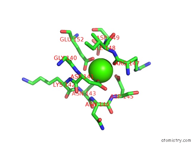
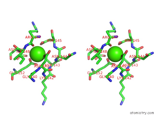
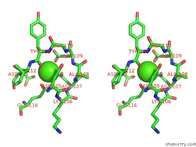
Calcium binding site 1 out of 6 in 1j1e
Go back to
Calcium binding site 1 out
of 6 in the Crystal Structure of the 52KDA Domain of Human Cardiac Troponin in the CA2+ Saturated Form
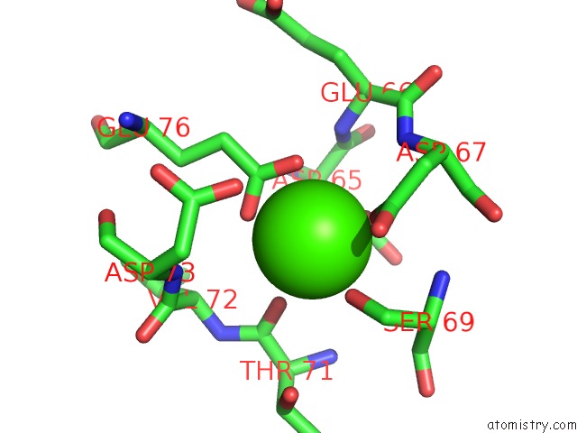
Mono view
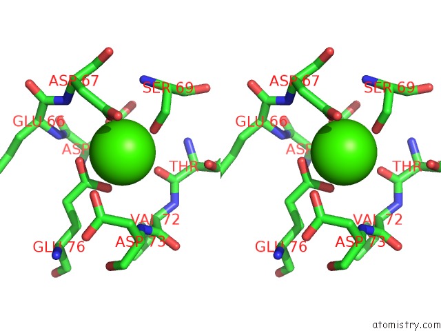
Stereo pair view
Mono view
Stereo pair view
A full contact list of Calcium with other atoms in the Ca binding
site number 1 of Crystal Structure of the 52KDA Domain of Human Cardiac Troponin in the CA2+ Saturated Form within 5.0Å range:
|
Calcium binding site 2 out of 6 in 1j1e
Go back to
Calcium binding site 2 out
of 6 in the Crystal Structure of the 52KDA Domain of Human Cardiac Troponin in the CA2+ Saturated Form
Mono view
Stereo pair view
Mono view
Stereo pair view
A full contact list of Calcium with other atoms in the Ca binding
site number 2 of Crystal Structure of the 52KDA Domain of Human Cardiac Troponin in the CA2+ Saturated Form within 5.0Å range:
|
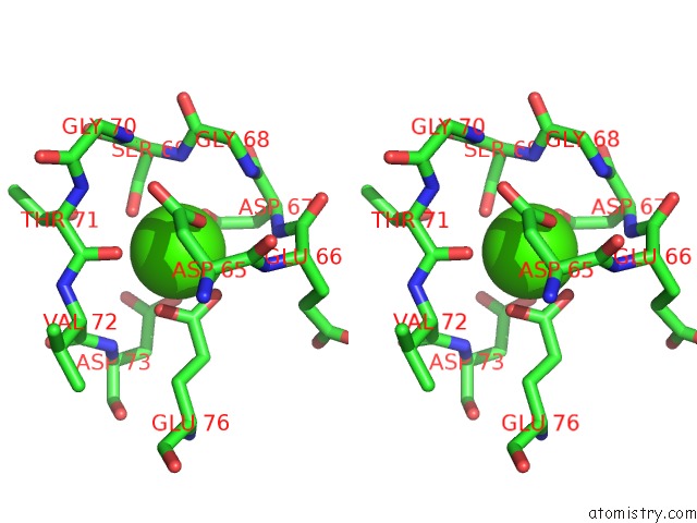
Calcium binding site 3 out of 6 in 1j1e
Go back to
Calcium binding site 3 out
of 6 in the Crystal Structure of the 52KDA Domain of Human Cardiac Troponin in the CA2+ Saturated Form
Mono view
Stereo pair view
Mono view
Stereo pair view
A full contact list of Calcium with other atoms in the Ca binding
site number 3 of Crystal Structure of the 52KDA Domain of Human Cardiac Troponin in the CA2+ Saturated Form within 5.0Å range:
|
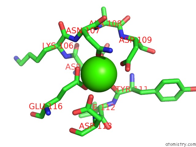
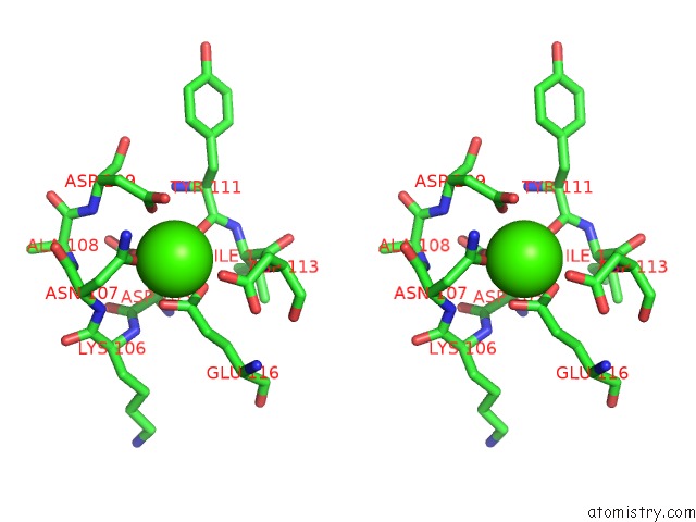
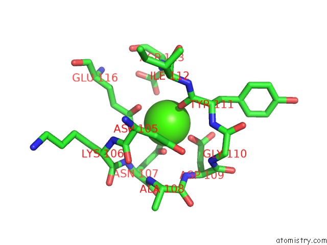
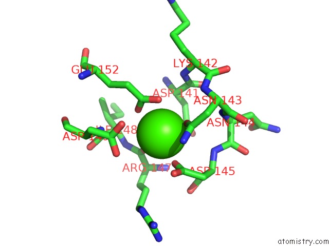
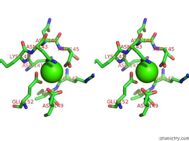
Calcium binding site 4 out of 6 in 1j1e
Go back to
Calcium binding site 4 out
of 6 in the Crystal Structure of the 52KDA Domain of Human Cardiac Troponin in the CA2+ Saturated Form

Mono view
Stereo pair view
Mono view
Stereo pair view
A full contact list of Calcium with other atoms in the Ca binding
site number 4 of Crystal Structure of the 52KDA Domain of Human Cardiac Troponin in the CA2+ Saturated Form within 5.0Å range:
|
Calcium binding site 5 out of 6 in 1j1e
Go back to
Calcium binding site 5 out
of 6 in the Crystal Structure of the 52KDA Domain of Human Cardiac Troponin in the CA2+ Saturated Form
Mono view
Stereo pair view
Mono view
Stereo pair view
A full contact list of Calcium with other atoms in the Ca binding
site number 5 of Crystal Structure of the 52KDA Domain of Human Cardiac Troponin in the CA2+ Saturated Form within 5.0Å range:
|
Calcium binding site 6 out of 6 in 1j1e
Go back to
Calcium binding site 6 out
of 6 in the Crystal Structure of the 52KDA Domain of Human Cardiac Troponin in the CA2+ Saturated Form
Mono view
Stereo pair view
Mono view
Stereo pair view
A full contact list of Calcium with other atoms in the Ca binding
site number 6 of Crystal Structure of the 52KDA Domain of Human Cardiac Troponin in the CA2+ Saturated Form within 5.0Å range:
|
Reference:
S.Takeda,
A.Yamashita,
K.Maeda,
Y.Maeda.
Structure of the Core Domain of Human Cardiac Troponin in the CA2+-Saturated Form Nature V. 424 35 2003.
ISSN: ISSN 0028-0836
PubMed: 12840750
DOI: 10.1038/NATURE01780
Page generated: Mon Jul 7 15:59:35 2025
ISSN: ISSN 0028-0836
PubMed: 12840750
DOI: 10.1038/NATURE01780
Last articles
Cl in 8BBVCl in 8BAJ
Cl in 8B7R
Cl in 8BAT
Cl in 8B9X
Cl in 8B9M
Cl in 8B9W
Cl in 8B8Z
Cl in 8B91
Cl in 8B90