Calcium »
PDB 1j1t-1jee »
1j34 »
Calcium in PDB 1j34: Crystal Structure of Mg(II)-and Ca(II)-Bound Gla Domain of Factor IX Complexed with Binding Protein
Enzymatic activity of Crystal Structure of Mg(II)-and Ca(II)-Bound Gla Domain of Factor IX Complexed with Binding Protein
All present enzymatic activity of Crystal Structure of Mg(II)-and Ca(II)-Bound Gla Domain of Factor IX Complexed with Binding Protein:
3.4.21.22;
3.4.21.22;
Protein crystallography data
The structure of Crystal Structure of Mg(II)-and Ca(II)-Bound Gla Domain of Factor IX Complexed with Binding Protein, PDB code: 1j34
was solved by
Y.Shikamoto,
T.Morita,
Z.Fujimoto,
H.Mizuno,
with X-Ray Crystallography technique. A brief refinement statistics is given in the table below:
| Resolution Low / High (Å) | 19.63 / 1.55 |
| Space group | C 1 2 1 |
| Cell size a, b, c (Å), α, β, γ (°) | 128.552, 37.204, 62.626, 90.00, 103.56, 90.00 |
| R / Rfree (%) | 18.3 / 21.2 |
Other elements in 1j34:
The structure of Crystal Structure of Mg(II)-and Ca(II)-Bound Gla Domain of Factor IX Complexed with Binding Protein also contains other interesting chemical elements:
| Magnesium | (Mg) | 3 atoms |
Calcium Binding Sites:
The binding sites of Calcium atom in the Crystal Structure of Mg(II)-and Ca(II)-Bound Gla Domain of Factor IX Complexed with Binding Protein
(pdb code 1j34). This binding sites where shown within
5.0 Angstroms radius around Calcium atom.
In total 7 binding sites of Calcium where determined in the Crystal Structure of Mg(II)-and Ca(II)-Bound Gla Domain of Factor IX Complexed with Binding Protein, PDB code: 1j34:
Jump to Calcium binding site number: 1; 2; 3; 4; 5; 6; 7;
In total 7 binding sites of Calcium where determined in the Crystal Structure of Mg(II)-and Ca(II)-Bound Gla Domain of Factor IX Complexed with Binding Protein, PDB code: 1j34:
Jump to Calcium binding site number: 1; 2; 3; 4; 5; 6; 7;
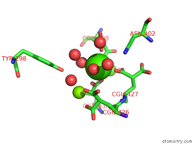
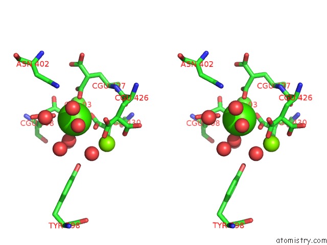
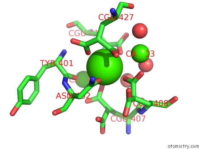
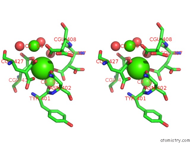
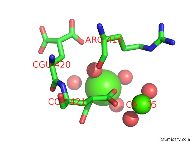
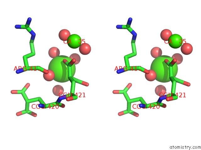
Calcium binding site 1 out of 7 in 1j34
Go back to
Calcium binding site 1 out
of 7 in the Crystal Structure of Mg(II)-and Ca(II)-Bound Gla Domain of Factor IX Complexed with Binding Protein
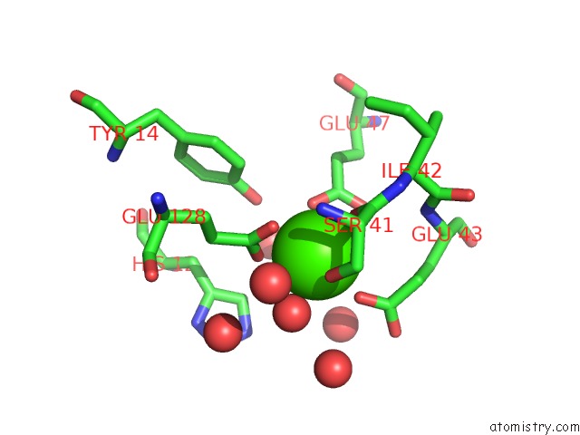
Mono view
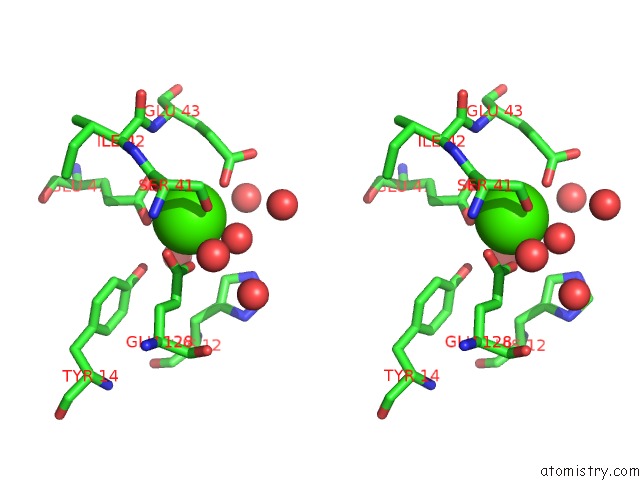
Stereo pair view
Mono view
Stereo pair view
A full contact list of Calcium with other atoms in the Ca binding
site number 1 of Crystal Structure of Mg(II)-and Ca(II)-Bound Gla Domain of Factor IX Complexed with Binding Protein within 5.0Å range:
|
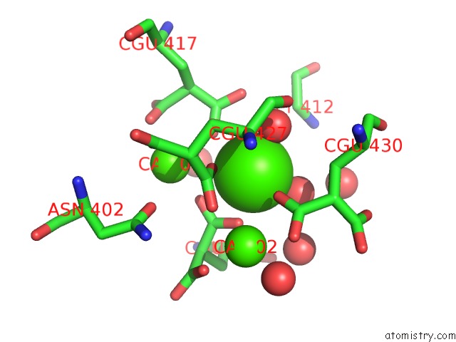
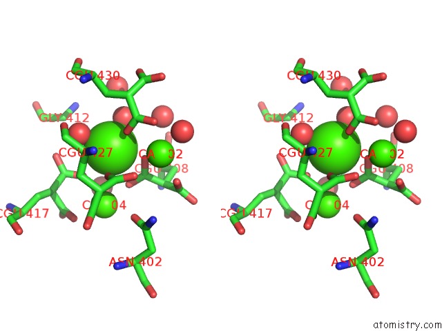
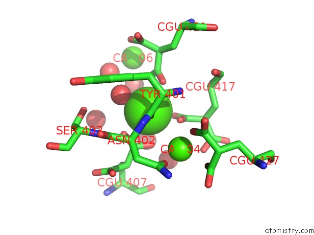
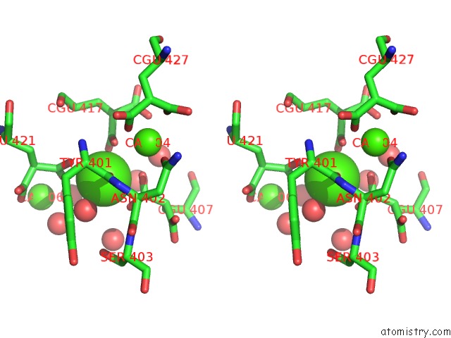
Calcium binding site 2 out of 7 in 1j34
Go back to
Calcium binding site 2 out
of 7 in the Crystal Structure of Mg(II)-and Ca(II)-Bound Gla Domain of Factor IX Complexed with Binding Protein
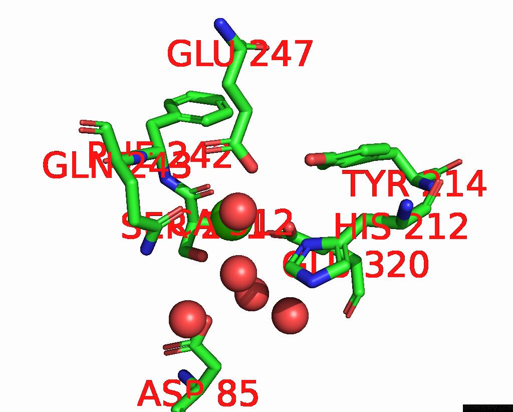
Mono view
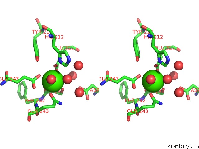
Stereo pair view
Mono view
Stereo pair view
A full contact list of Calcium with other atoms in the Ca binding
site number 2 of Crystal Structure of Mg(II)-and Ca(II)-Bound Gla Domain of Factor IX Complexed with Binding Protein within 5.0Å range:
|
Calcium binding site 3 out of 7 in 1j34
Go back to
Calcium binding site 3 out
of 7 in the Crystal Structure of Mg(II)-and Ca(II)-Bound Gla Domain of Factor IX Complexed with Binding Protein
Mono view
Stereo pair view
Mono view
Stereo pair view
A full contact list of Calcium with other atoms in the Ca binding
site number 3 of Crystal Structure of Mg(II)-and Ca(II)-Bound Gla Domain of Factor IX Complexed with Binding Protein within 5.0Å range:
|
Calcium binding site 4 out of 7 in 1j34
Go back to
Calcium binding site 4 out
of 7 in the Crystal Structure of Mg(II)-and Ca(II)-Bound Gla Domain of Factor IX Complexed with Binding Protein
Mono view
Stereo pair view
Mono view
Stereo pair view
A full contact list of Calcium with other atoms in the Ca binding
site number 4 of Crystal Structure of Mg(II)-and Ca(II)-Bound Gla Domain of Factor IX Complexed with Binding Protein within 5.0Å range:
|
Calcium binding site 5 out of 7 in 1j34
Go back to
Calcium binding site 5 out
of 7 in the Crystal Structure of Mg(II)-and Ca(II)-Bound Gla Domain of Factor IX Complexed with Binding Protein
Mono view
Stereo pair view
Mono view
Stereo pair view
A full contact list of Calcium with other atoms in the Ca binding
site number 5 of Crystal Structure of Mg(II)-and Ca(II)-Bound Gla Domain of Factor IX Complexed with Binding Protein within 5.0Å range:
|
Calcium binding site 6 out of 7 in 1j34
Go back to
Calcium binding site 6 out
of 7 in the Crystal Structure of Mg(II)-and Ca(II)-Bound Gla Domain of Factor IX Complexed with Binding Protein
Mono view
Stereo pair view
Mono view
Stereo pair view
A full contact list of Calcium with other atoms in the Ca binding
site number 6 of Crystal Structure of Mg(II)-and Ca(II)-Bound Gla Domain of Factor IX Complexed with Binding Protein within 5.0Å range:
|
Calcium binding site 7 out of 7 in 1j34
Go back to
Calcium binding site 7 out
of 7 in the Crystal Structure of Mg(II)-and Ca(II)-Bound Gla Domain of Factor IX Complexed with Binding Protein
Mono view
Stereo pair view
Mono view
Stereo pair view
A full contact list of Calcium with other atoms in the Ca binding
site number 7 of Crystal Structure of Mg(II)-and Ca(II)-Bound Gla Domain of Factor IX Complexed with Binding Protein within 5.0Å range:
|
Reference:
Y.Shikamoto,
T.Morita,
Z.Fujimoto,
H.Mizuno.
Crystal Structure of MG2+- and CA2+-Bound Gla Domain of Factor IX Complexed with Binding Protein J.Biol.Chem. V. 278 24090 2003.
ISSN: ISSN 0021-9258
PubMed: 12695512
DOI: 10.1074/JBC.M300650200
Page generated: Mon Jul 7 16:00:54 2025
ISSN: ISSN 0021-9258
PubMed: 12695512
DOI: 10.1074/JBC.M300650200
Last articles
Cl in 5JENCl in 5JFD
Cl in 5JEU
Cl in 5JEC
Cl in 5JDT
Cl in 5JED
Cl in 5JD4
Cl in 5JDS
Cl in 5JDU
Cl in 5JD3