Calcium »
PDB 1j1t-1jee »
1j55 »
Calcium in PDB 1j55: The Crystal Structure of Ca+-Bound Human S100P Determined at 2.0A Resolution By X-Ray
Protein crystallography data
The structure of The Crystal Structure of Ca+-Bound Human S100P Determined at 2.0A Resolution By X-Ray, PDB code: 1j55
was solved by
H.Zhang,
G.Wang,
Y.Ding,
Z.Wang,
R.Barraclough,
P.S.Rudland,
D.G.Fernig,
Z.Rao,
with X-Ray Crystallography technique. A brief refinement statistics is given in the table below:
| Resolution Low / High (Å) | 40.00 / 2.00 |
| Space group | P 41 21 2 |
| Cell size a, b, c (Å), α, β, γ (°) | 60.827, 60.827, 47.610, 90.00, 90.00, 90.00 |
| R / Rfree (%) | 21.4 / 26.7 |
Calcium Binding Sites:
The binding sites of Calcium atom in the The Crystal Structure of Ca+-Bound Human S100P Determined at 2.0A Resolution By X-Ray
(pdb code 1j55). This binding sites where shown within
5.0 Angstroms radius around Calcium atom.
In total 2 binding sites of Calcium where determined in the The Crystal Structure of Ca+-Bound Human S100P Determined at 2.0A Resolution By X-Ray, PDB code: 1j55:
Jump to Calcium binding site number: 1; 2;
In total 2 binding sites of Calcium where determined in the The Crystal Structure of Ca+-Bound Human S100P Determined at 2.0A Resolution By X-Ray, PDB code: 1j55:
Jump to Calcium binding site number: 1; 2;
Calcium binding site 1 out of 2 in 1j55
Go back to
Calcium binding site 1 out
of 2 in the The Crystal Structure of Ca+-Bound Human S100P Determined at 2.0A Resolution By X-Ray
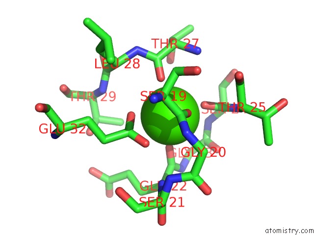
Mono view
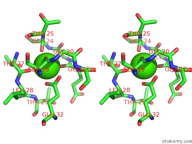
Stereo pair view
Mono view
Stereo pair view
A full contact list of Calcium with other atoms in the Ca binding
site number 1 of The Crystal Structure of Ca+-Bound Human S100P Determined at 2.0A Resolution By X-Ray within 5.0Å range:
|
Calcium binding site 2 out of 2 in 1j55
Go back to
Calcium binding site 2 out
of 2 in the The Crystal Structure of Ca+-Bound Human S100P Determined at 2.0A Resolution By X-Ray
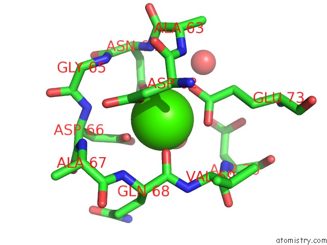
Mono view
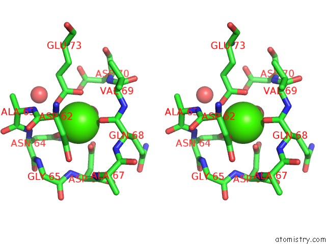
Stereo pair view
Mono view
Stereo pair view
A full contact list of Calcium with other atoms in the Ca binding
site number 2 of The Crystal Structure of Ca+-Bound Human S100P Determined at 2.0A Resolution By X-Ray within 5.0Å range:
|
Reference:
H.Zhang,
G.Wang,
Y.Ding,
Z.Wang,
R.Barraclough,
P.S.Rudland,
D.G.Fernig,
Z.Rao.
The Crystal Structure at 2A Resolution of the CA2+-Binding Protein S100P J.Mol.Biol. V. 325 785 2003.
ISSN: ISSN 0022-2836
PubMed: 12507480
DOI: 10.1016/S0022-2836(02)01278-0
Page generated: Thu Jul 11 10:36:07 2024
ISSN: ISSN 0022-2836
PubMed: 12507480
DOI: 10.1016/S0022-2836(02)01278-0
Last articles
Zn in 9MJ5Zn in 9HNW
Zn in 9G0L
Zn in 9FNE
Zn in 9DZN
Zn in 9E0I
Zn in 9D32
Zn in 9DAK
Zn in 8ZXC
Zn in 8ZUF