Calcium »
PDB 1j1t-1jee »
1j8a »
Calcium in PDB 1j8a: Crystal Structure of Benzamidine Inhibited Bovine Pancreatic Trypsin at 105K to 1.21A Resolution From Laboratory Source with High Number of Waters Modelled
Enzymatic activity of Crystal Structure of Benzamidine Inhibited Bovine Pancreatic Trypsin at 105K to 1.21A Resolution From Laboratory Source with High Number of Waters Modelled
All present enzymatic activity of Crystal Structure of Benzamidine Inhibited Bovine Pancreatic Trypsin at 105K to 1.21A Resolution From Laboratory Source with High Number of Waters Modelled:
3.4.21.4;
3.4.21.4;
Protein crystallography data
The structure of Crystal Structure of Benzamidine Inhibited Bovine Pancreatic Trypsin at 105K to 1.21A Resolution From Laboratory Source with High Number of Waters Modelled, PDB code: 1j8a
was solved by
J.A.Cuesta-Seijo,
S.Garcia-Granda,
with X-Ray Crystallography technique. A brief refinement statistics is given in the table below:
| Resolution Low / High (Å) | 19.90 / 1.21 |
| Space group | P 21 21 21 |
| Cell size a, b, c (Å), α, β, γ (°) | 54.275, 58.568, 66.141, 90.00, 90.00, 90.00 |
| R / Rfree (%) | 16 / 17.8 |
Calcium Binding Sites:
The binding sites of Calcium atom in the Crystal Structure of Benzamidine Inhibited Bovine Pancreatic Trypsin at 105K to 1.21A Resolution From Laboratory Source with High Number of Waters Modelled
(pdb code 1j8a). This binding sites where shown within
5.0 Angstroms radius around Calcium atom.
In total only one binding site of Calcium was determined in the Crystal Structure of Benzamidine Inhibited Bovine Pancreatic Trypsin at 105K to 1.21A Resolution From Laboratory Source with High Number of Waters Modelled, PDB code: 1j8a:
In total only one binding site of Calcium was determined in the Crystal Structure of Benzamidine Inhibited Bovine Pancreatic Trypsin at 105K to 1.21A Resolution From Laboratory Source with High Number of Waters Modelled, PDB code: 1j8a:
Calcium binding site 1 out of 1 in 1j8a
Go back to
Calcium binding site 1 out
of 1 in the Crystal Structure of Benzamidine Inhibited Bovine Pancreatic Trypsin at 105K to 1.21A Resolution From Laboratory Source with High Number of Waters Modelled
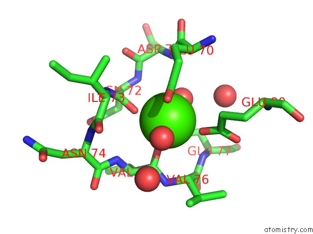
Mono view
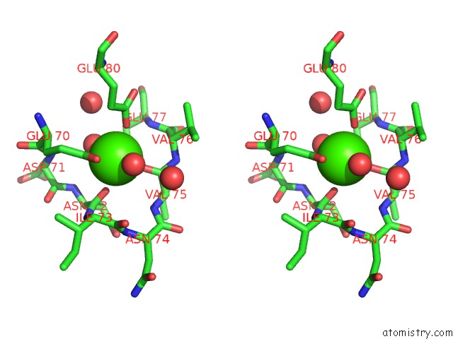
Stereo pair view
Mono view
Stereo pair view
A full contact list of Calcium with other atoms in the Ca binding
site number 1 of Crystal Structure of Benzamidine Inhibited Bovine Pancreatic Trypsin at 105K to 1.21A Resolution From Laboratory Source with High Number of Waters Modelled within 5.0Å range:
|
Reference:
J.A.Cuesta-Seijo,
S.Garcia-Granda.
Trypsin As A Model For High Resolution X-Ray Diffraction in Proteins. Bol.R.Soc.Hist.Nat.Sec.Geol. V. 97 123 2002.
ISSN: ISSN 5083-7510
Page generated: Thu Jul 11 10:38:31 2024
ISSN: ISSN 5083-7510
Last articles
Zn in 9MJ5Zn in 9HNW
Zn in 9G0L
Zn in 9FNE
Zn in 9DZN
Zn in 9E0I
Zn in 9D32
Zn in 9DAK
Zn in 8ZXC
Zn in 8ZUF