Calcium »
PDB 1j1t-1jee »
1jap »
Calcium in PDB 1jap: Complex of Pro-Leu-Gly-Hydroxylamine with the Catalytic Domain of Matrix Metallo Proteinase-8 (MET80 Form)
Enzymatic activity of Complex of Pro-Leu-Gly-Hydroxylamine with the Catalytic Domain of Matrix Metallo Proteinase-8 (MET80 Form)
All present enzymatic activity of Complex of Pro-Leu-Gly-Hydroxylamine with the Catalytic Domain of Matrix Metallo Proteinase-8 (MET80 Form):
3.4.24.34;
3.4.24.34;
Protein crystallography data
The structure of Complex of Pro-Leu-Gly-Hydroxylamine with the Catalytic Domain of Matrix Metallo Proteinase-8 (MET80 Form), PDB code: 1jap
was solved by
W.Bode,
P.Reinemer,
R.Huber,
T.Kleine,
S.Schnierer,
H.Tschesche,
with X-Ray Crystallography technique. A brief refinement statistics is given in the table below:
| Resolution Low / High (Å) | 6.00 / 1.82 |
| Space group | P 21 21 21 |
| Cell size a, b, c (Å), α, β, γ (°) | 33.090, 69.370, 72.480, 90.00, 90.00, 90.00 |
| R / Rfree (%) | 19.4 / n/a |
Other elements in 1jap:
The structure of Complex of Pro-Leu-Gly-Hydroxylamine with the Catalytic Domain of Matrix Metallo Proteinase-8 (MET80 Form) also contains other interesting chemical elements:
| Zinc | (Zn) | 2 atoms |
Calcium Binding Sites:
The binding sites of Calcium atom in the Complex of Pro-Leu-Gly-Hydroxylamine with the Catalytic Domain of Matrix Metallo Proteinase-8 (MET80 Form)
(pdb code 1jap). This binding sites where shown within
5.0 Angstroms radius around Calcium atom.
In total 2 binding sites of Calcium where determined in the Complex of Pro-Leu-Gly-Hydroxylamine with the Catalytic Domain of Matrix Metallo Proteinase-8 (MET80 Form), PDB code: 1jap:
Jump to Calcium binding site number: 1; 2;
In total 2 binding sites of Calcium where determined in the Complex of Pro-Leu-Gly-Hydroxylamine with the Catalytic Domain of Matrix Metallo Proteinase-8 (MET80 Form), PDB code: 1jap:
Jump to Calcium binding site number: 1; 2;
Calcium binding site 1 out of 2 in 1jap
Go back to
Calcium binding site 1 out
of 2 in the Complex of Pro-Leu-Gly-Hydroxylamine with the Catalytic Domain of Matrix Metallo Proteinase-8 (MET80 Form)
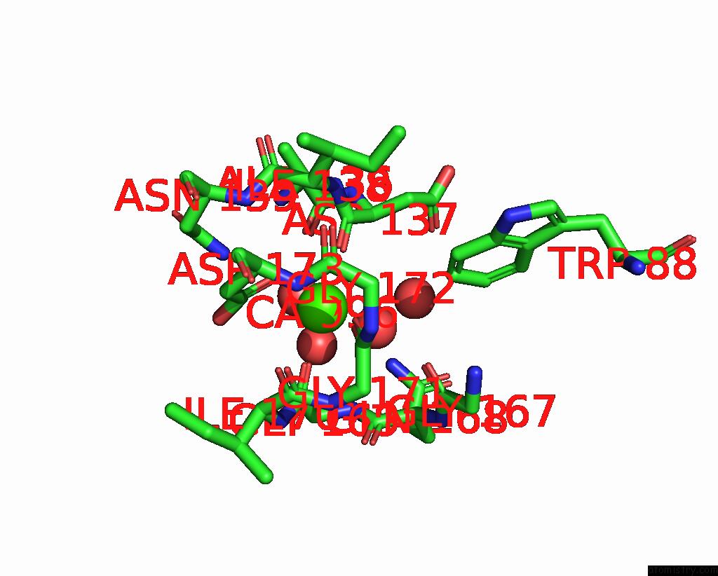
Mono view
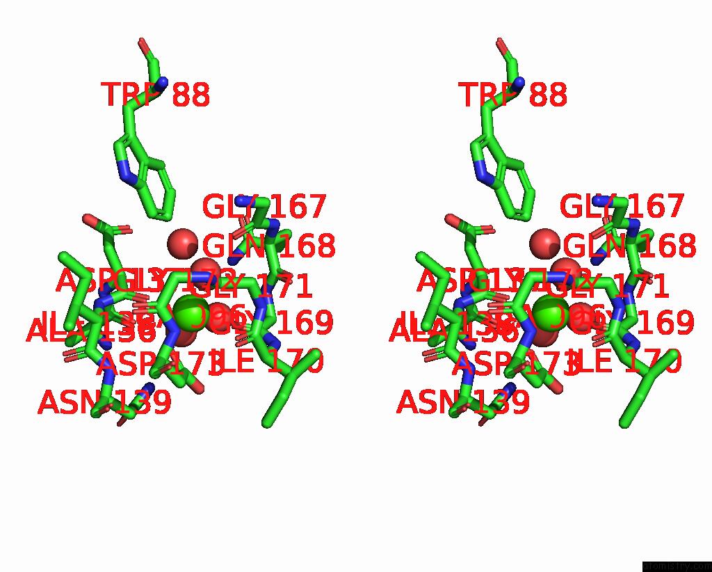
Stereo pair view
Mono view
Stereo pair view
A full contact list of Calcium with other atoms in the Ca binding
site number 1 of Complex of Pro-Leu-Gly-Hydroxylamine with the Catalytic Domain of Matrix Metallo Proteinase-8 (MET80 Form) within 5.0Å range:
|
Calcium binding site 2 out of 2 in 1jap
Go back to
Calcium binding site 2 out
of 2 in the Complex of Pro-Leu-Gly-Hydroxylamine with the Catalytic Domain of Matrix Metallo Proteinase-8 (MET80 Form)
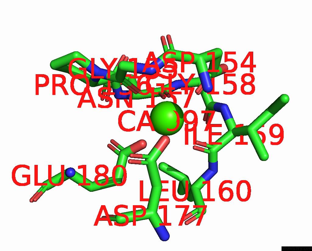
Mono view
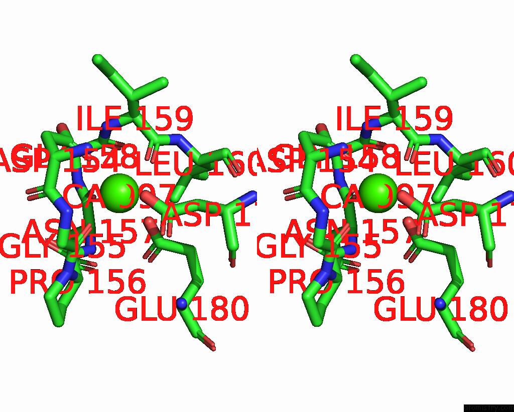
Stereo pair view
Mono view
Stereo pair view
A full contact list of Calcium with other atoms in the Ca binding
site number 2 of Complex of Pro-Leu-Gly-Hydroxylamine with the Catalytic Domain of Matrix Metallo Proteinase-8 (MET80 Form) within 5.0Å range:
|
Reference:
W.Bode,
P.Reinemer,
R.Huber,
T.Kleine,
S.Schnierer,
H.Tschesche.
The X-Ray Crystal Structure of the Catalytic Domain of Human Neutrophil Collagenase Inhibited By A Substrate Analogue Reveals the Essentials For Catalysis and Specificity. Embo J. V. 13 1263 1994.
ISSN: ISSN 0261-4189
PubMed: 8137810
Page generated: Mon Jul 7 16:04:38 2025
ISSN: ISSN 0261-4189
PubMed: 8137810
Last articles
Fe in 2YXOFe in 2YRS
Fe in 2YXC
Fe in 2YNM
Fe in 2YVJ
Fe in 2YP1
Fe in 2YU2
Fe in 2YU1
Fe in 2YQB
Fe in 2YOO