Calcium »
PDB 1j1t-1jee »
1je5 »
Calcium in PDB 1je5: Crystal Structure of GP2.5, A Single-Stranded Dna Binding Protein Encoded By Bacteriophage T7
Protein crystallography data
The structure of Crystal Structure of GP2.5, A Single-Stranded Dna Binding Protein Encoded By Bacteriophage T7, PDB code: 1je5
was solved by
T.Hollis,
J.M.Stattel,
D.S.Walther,
C.C.Richardson,
T.E.Ellenberger,
with X-Ray Crystallography technique. A brief refinement statistics is given in the table below:
| Resolution Low / High (Å) | 24.71 / 1.90 |
| Space group | P 21 21 21 |
| Cell size a, b, c (Å), α, β, γ (°) | 68.100, 71.800, 82.200, 90.00, 90.00, 90.00 |
| R / Rfree (%) | 22.1 / 26.8 |
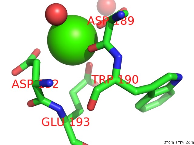
Calcium Binding Sites:
The binding sites of Calcium atom in the Crystal Structure of GP2.5, A Single-Stranded Dna Binding Protein Encoded By Bacteriophage T7
(pdb code 1je5). This binding sites where shown within
5.0 Angstroms radius around Calcium atom.
In total only one binding site of Calcium was determined in the Crystal Structure of GP2.5, A Single-Stranded Dna Binding Protein Encoded By Bacteriophage T7, PDB code: 1je5:
In total only one binding site of Calcium was determined in the Crystal Structure of GP2.5, A Single-Stranded Dna Binding Protein Encoded By Bacteriophage T7, PDB code: 1je5:
Calcium binding site 1 out of 1 in 1je5
Go back to
Calcium binding site 1 out
of 1 in the Crystal Structure of GP2.5, A Single-Stranded Dna Binding Protein Encoded By Bacteriophage T7
Mono view
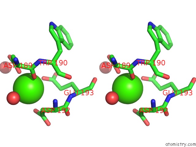
Stereo pair view
Mono view
Stereo pair view
A full contact list of Calcium with other atoms in the Ca binding
site number 1 of Crystal Structure of GP2.5, A Single-Stranded Dna Binding Protein Encoded By Bacteriophage T7 within 5.0Å range:
|
Reference:
T.Hollis,
J.M.Stattel,
D.S.Walther,
C.C.Richardson,
T.Ellenberger.
Structure of the Gene 2.5 Protein, A Single-Stranded Dna Binding Protein Encoded By Bacteriophage T7. Proc.Natl.Acad.Sci.Usa V. 98 9557 2001.
ISSN: ISSN 0027-8424
PubMed: 11481454
DOI: 10.1073/PNAS.171317698
Page generated: Thu Jul 11 10:43:18 2024
ISSN: ISSN 0027-8424
PubMed: 11481454
DOI: 10.1073/PNAS.171317698
Last articles
Zn in 9J0NZn in 9J0O
Zn in 9J0P
Zn in 9FJX
Zn in 9EKB
Zn in 9C0F
Zn in 9CAH
Zn in 9CH0
Zn in 9CH3
Zn in 9CH1