Calcium »
PDB 1j1t-1jee »
1jed »
Calcium in PDB 1jed: Crystal Structure of Atp Sulfurylase in Complex with Adp
Enzymatic activity of Crystal Structure of Atp Sulfurylase in Complex with Adp
All present enzymatic activity of Crystal Structure of Atp Sulfurylase in Complex with Adp:
2.7.7.4;
2.7.7.4;
Protein crystallography data
The structure of Crystal Structure of Atp Sulfurylase in Complex with Adp, PDB code: 1jed
was solved by
T.C.Ullrich,
R.Huber,
with X-Ray Crystallography technique. A brief refinement statistics is given in the table below:
| Resolution Low / High (Å) | 24.81 / 2.95 |
| Space group | H 3 2 |
| Cell size a, b, c (Å), α, β, γ (°) | 186.135, 186.135, 223.253, 90.00, 90.00, 120.00 |
| R / Rfree (%) | 19.2 / 22.7 |
Other elements in 1jed:
The structure of Crystal Structure of Atp Sulfurylase in Complex with Adp also contains other interesting chemical elements:
| Cadmium | (Cd) | 11 atoms |
| Magnesium | (Mg) | 4 atoms |
| Sodium | (Na) | 12 atoms |
Calcium Binding Sites:
The binding sites of Calcium atom in the Crystal Structure of Atp Sulfurylase in Complex with Adp
(pdb code 1jed). This binding sites where shown within
5.0 Angstroms radius around Calcium atom.
In total 6 binding sites of Calcium where determined in the Crystal Structure of Atp Sulfurylase in Complex with Adp, PDB code: 1jed:
Jump to Calcium binding site number: 1; 2; 3; 4; 5; 6;
In total 6 binding sites of Calcium where determined in the Crystal Structure of Atp Sulfurylase in Complex with Adp, PDB code: 1jed:
Jump to Calcium binding site number: 1; 2; 3; 4; 5; 6;
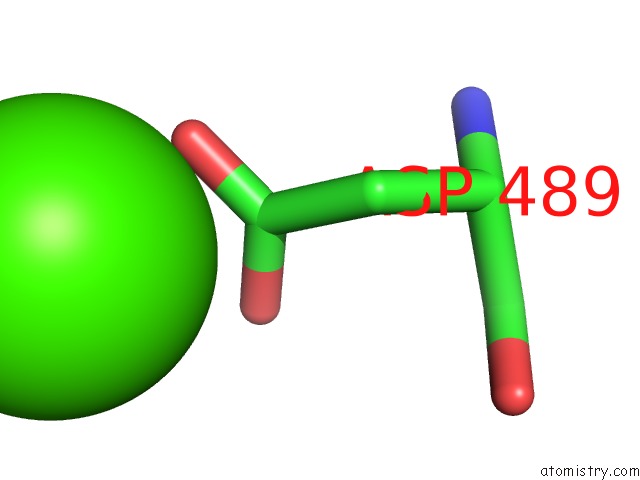
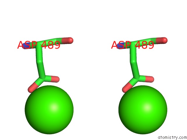
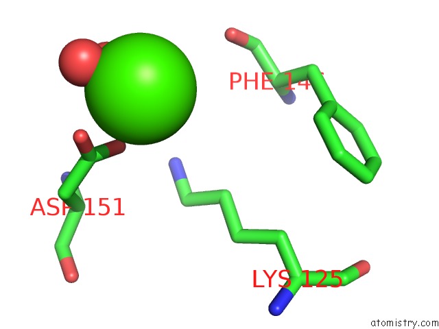
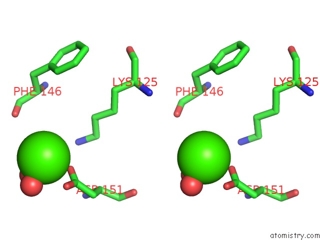
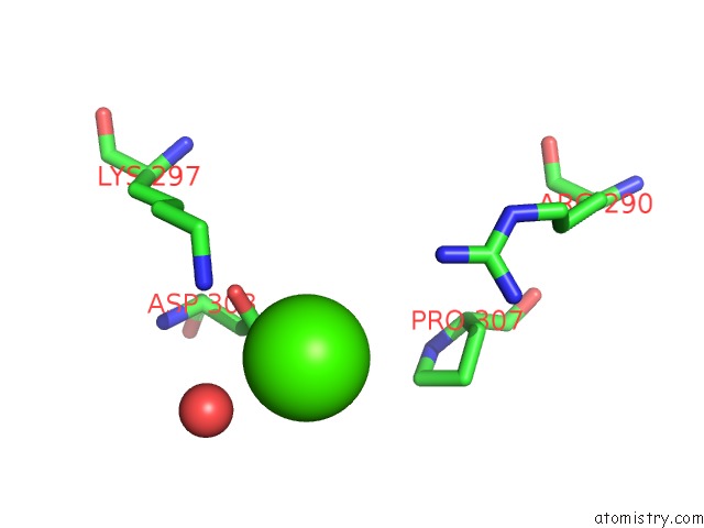
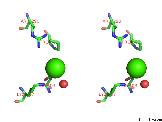
Calcium binding site 1 out of 6 in 1jed
Go back to
Calcium binding site 1 out
of 6 in the Crystal Structure of Atp Sulfurylase in Complex with Adp
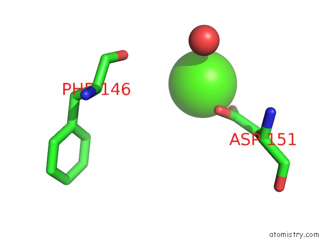
Mono view
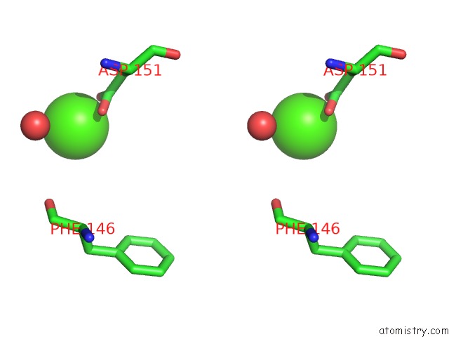
Stereo pair view
Mono view
Stereo pair view
A full contact list of Calcium with other atoms in the Ca binding
site number 1 of Crystal Structure of Atp Sulfurylase in Complex with Adp within 5.0Å range:
|
Calcium binding site 2 out of 6 in 1jed
Go back to
Calcium binding site 2 out
of 6 in the Crystal Structure of Atp Sulfurylase in Complex with Adp
Mono view
Stereo pair view
Mono view
Stereo pair view
A full contact list of Calcium with other atoms in the Ca binding
site number 2 of Crystal Structure of Atp Sulfurylase in Complex with Adp within 5.0Å range:
|
Calcium binding site 3 out of 6 in 1jed
Go back to
Calcium binding site 3 out
of 6 in the Crystal Structure of Atp Sulfurylase in Complex with Adp
Mono view
Stereo pair view
Mono view
Stereo pair view
A full contact list of Calcium with other atoms in the Ca binding
site number 3 of Crystal Structure of Atp Sulfurylase in Complex with Adp within 5.0Å range:
|
Calcium binding site 4 out of 6 in 1jed
Go back to
Calcium binding site 4 out
of 6 in the Crystal Structure of Atp Sulfurylase in Complex with Adp
Mono view
Stereo pair view
Mono view
Stereo pair view
A full contact list of Calcium with other atoms in the Ca binding
site number 4 of Crystal Structure of Atp Sulfurylase in Complex with Adp within 5.0Å range:
|
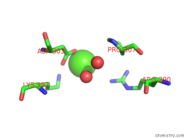
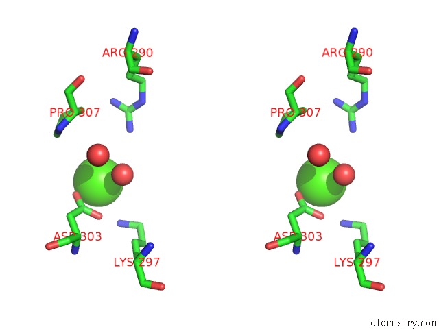
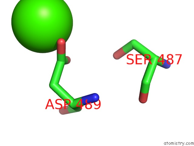
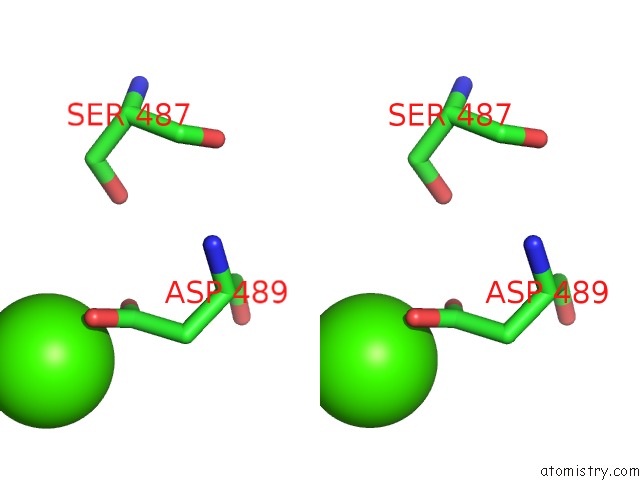
Calcium binding site 5 out of 6 in 1jed
Go back to
Calcium binding site 5 out
of 6 in the Crystal Structure of Atp Sulfurylase in Complex with Adp
Mono view
Stereo pair view
Mono view
Stereo pair view
A full contact list of Calcium with other atoms in the Ca binding
site number 5 of Crystal Structure of Atp Sulfurylase in Complex with Adp within 5.0Å range:
|
Calcium binding site 6 out of 6 in 1jed
Go back to
Calcium binding site 6 out
of 6 in the Crystal Structure of Atp Sulfurylase in Complex with Adp
Mono view
Stereo pair view
Mono view
Stereo pair view
A full contact list of Calcium with other atoms in the Ca binding
site number 6 of Crystal Structure of Atp Sulfurylase in Complex with Adp within 5.0Å range:
|
Reference:
T.C.Ullrich,
R.Huber.
The Complex Structures of Atp Sulfurylase with Thiosulfate, Adp and Chlorate Reveal New Insights in Inhibitory Effects and the Catalytic Cycle. J.Mol.Biol. V. 313 1117 2001.
ISSN: ISSN 0022-2836
PubMed: 11700067
DOI: 10.1006/JMBI.2001.5098
Page generated: Thu Jul 11 10:43:53 2024
ISSN: ISSN 0022-2836
PubMed: 11700067
DOI: 10.1006/JMBI.2001.5098
Last articles
Zn in 9MJ5Zn in 9HNW
Zn in 9G0L
Zn in 9FNE
Zn in 9DZN
Zn in 9E0I
Zn in 9D32
Zn in 9DAK
Zn in 8ZXC
Zn in 8ZUF