Calcium »
PDB 1jf5-1jv2 »
1jix »
Calcium in PDB 1jix: T4 Phage Bgt in Complex with CA2+
Enzymatic activity of T4 Phage Bgt in Complex with CA2+
All present enzymatic activity of T4 Phage Bgt in Complex with CA2+:
2.4.1.27;
2.4.1.27;
Protein crystallography data
The structure of T4 Phage Bgt in Complex with CA2+, PDB code: 1jix
was solved by
S.Morera,
L.Lariviere,
J.Kurzeck,
U.Aschke-Sonnenborn,
P.S.Freemont,
J.Janin,
W.Ruger,
with X-Ray Crystallography technique. A brief refinement statistics is given in the table below:
| Resolution Low / High (Å) | 20.00 / 1.65 |
| Space group | P 1 21 1 |
| Cell size a, b, c (Å), α, β, γ (°) | 47.943, 71.454, 62.010, 90.00, 91.36, 90.00 |
| R / Rfree (%) | 20 / 21.4 |
Calcium Binding Sites:
The binding sites of Calcium atom in the T4 Phage Bgt in Complex with CA2+
(pdb code 1jix). This binding sites where shown within
5.0 Angstroms radius around Calcium atom.
In total only one binding site of Calcium was determined in the T4 Phage Bgt in Complex with CA2+, PDB code: 1jix:
In total only one binding site of Calcium was determined in the T4 Phage Bgt in Complex with CA2+, PDB code: 1jix:
Calcium binding site 1 out of 1 in 1jix
Go back to
Calcium binding site 1 out
of 1 in the T4 Phage Bgt in Complex with CA2+
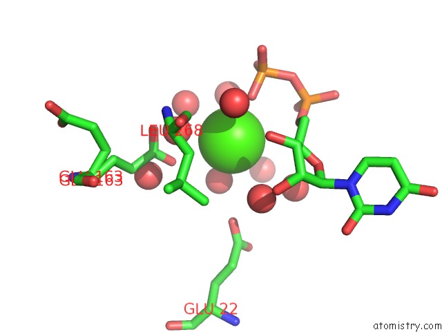
Mono view
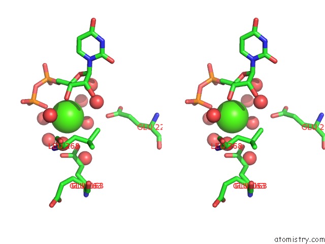
Stereo pair view
Mono view
Stereo pair view
A full contact list of Calcium with other atoms in the Ca binding
site number 1 of T4 Phage Bgt in Complex with CA2+ within 5.0Å range:
|
Reference:
S.Morera,
L.Lariviere,
J.Kurzeck,
U.Aschke-Sonnenborn,
P.S.Freemont,
J.Janin,
W.Ruger.
High Resolution Crystal Structures of T4 Phage Beta-Glucosyltransferase: Induced Fit and Effect of Substrate and Metal Binding. J.Mol.Biol. V. 311 569 2001.
ISSN: ISSN 0022-2836
PubMed: 11493010
DOI: 10.1006/JMBI.2001.4905
Page generated: Thu Jul 11 10:47:35 2024
ISSN: ISSN 0022-2836
PubMed: 11493010
DOI: 10.1006/JMBI.2001.4905
Last articles
Zn in 9MJ5Zn in 9HNW
Zn in 9G0L
Zn in 9FNE
Zn in 9DZN
Zn in 9E0I
Zn in 9D32
Zn in 9DAK
Zn in 8ZXC
Zn in 8ZUF