Calcium »
PDB 1jf5-1jv2 »
1jrq »
Calcium in PDB 1jrq: X-Ray Structure Analysis of the Role of the Conserved Tyrosine-369 in Active Site of E. Coli Amine Oxidase
Enzymatic activity of X-Ray Structure Analysis of the Role of the Conserved Tyrosine-369 in Active Site of E. Coli Amine Oxidase
All present enzymatic activity of X-Ray Structure Analysis of the Role of the Conserved Tyrosine-369 in Active Site of E. Coli Amine Oxidase:
1.4.3.4;
1.4.3.4;
Protein crystallography data
The structure of X-Ray Structure Analysis of the Role of the Conserved Tyrosine-369 in Active Site of E. Coli Amine Oxidase, PDB code: 1jrq
was solved by
J.M.Murray,
C.R.Kurtis,
W.Tambarajah,
C.G.Saysell,
C.M.Wilmot,
M.R.Parsons,
S.E.V.Phillips,
P.F.Knowles,
M.J.Mcpherson,
with X-Ray Crystallography technique. A brief refinement statistics is given in the table below:
| Resolution Low / High (Å) | 20.00 / 2.15 |
| Space group | P 21 21 21 |
| Cell size a, b, c (Å), α, β, γ (°) | 134.540, 166.450, 79.380, 90.00, 90.00, 90.00 |
| R / Rfree (%) | 19.5 / 23.5 |
Other elements in 1jrq:
The structure of X-Ray Structure Analysis of the Role of the Conserved Tyrosine-369 in Active Site of E. Coli Amine Oxidase also contains other interesting chemical elements:
| Copper | (Cu) | 2 atoms |
Calcium Binding Sites:
The binding sites of Calcium atom in the X-Ray Structure Analysis of the Role of the Conserved Tyrosine-369 in Active Site of E. Coli Amine Oxidase
(pdb code 1jrq). This binding sites where shown within
5.0 Angstroms radius around Calcium atom.
In total 4 binding sites of Calcium where determined in the X-Ray Structure Analysis of the Role of the Conserved Tyrosine-369 in Active Site of E. Coli Amine Oxidase, PDB code: 1jrq:
Jump to Calcium binding site number: 1; 2; 3; 4;
In total 4 binding sites of Calcium where determined in the X-Ray Structure Analysis of the Role of the Conserved Tyrosine-369 in Active Site of E. Coli Amine Oxidase, PDB code: 1jrq:
Jump to Calcium binding site number: 1; 2; 3; 4;
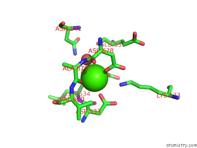
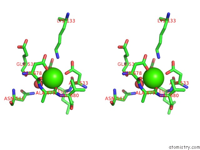
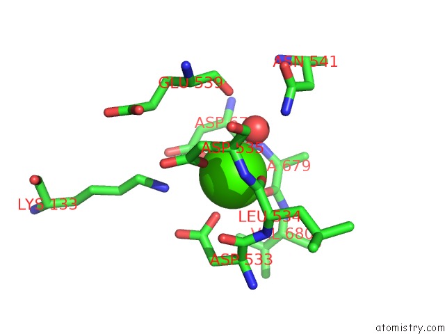
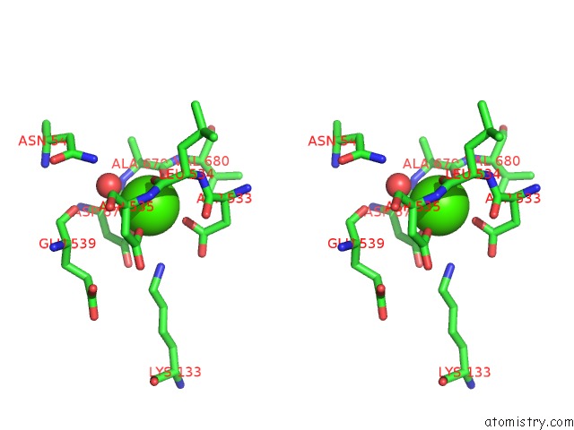
Calcium binding site 1 out of 4 in 1jrq
Go back to
Calcium binding site 1 out
of 4 in the X-Ray Structure Analysis of the Role of the Conserved Tyrosine-369 in Active Site of E. Coli Amine Oxidase
Mono view
Stereo pair view
Mono view
Stereo pair view
A full contact list of Calcium with other atoms in the Ca binding
site number 1 of X-Ray Structure Analysis of the Role of the Conserved Tyrosine-369 in Active Site of E. Coli Amine Oxidase within 5.0Å range:
|
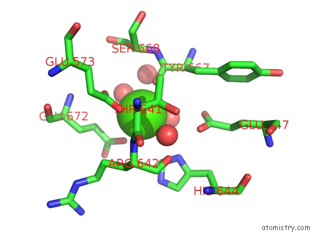
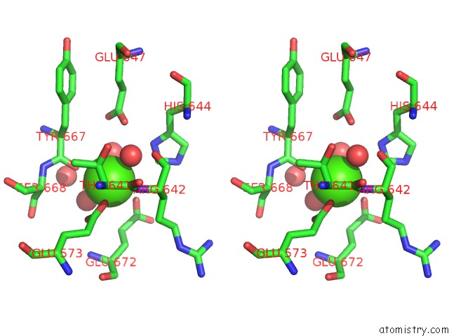
Calcium binding site 2 out of 4 in 1jrq
Go back to
Calcium binding site 2 out
of 4 in the X-Ray Structure Analysis of the Role of the Conserved Tyrosine-369 in Active Site of E. Coli Amine Oxidase
Mono view
Stereo pair view
Mono view
Stereo pair view
A full contact list of Calcium with other atoms in the Ca binding
site number 2 of X-Ray Structure Analysis of the Role of the Conserved Tyrosine-369 in Active Site of E. Coli Amine Oxidase within 5.0Å range:
|
Calcium binding site 3 out of 4 in 1jrq
Go back to
Calcium binding site 3 out
of 4 in the X-Ray Structure Analysis of the Role of the Conserved Tyrosine-369 in Active Site of E. Coli Amine Oxidase
Mono view
Stereo pair view
Mono view
Stereo pair view
A full contact list of Calcium with other atoms in the Ca binding
site number 3 of X-Ray Structure Analysis of the Role of the Conserved Tyrosine-369 in Active Site of E. Coli Amine Oxidase within 5.0Å range:
|
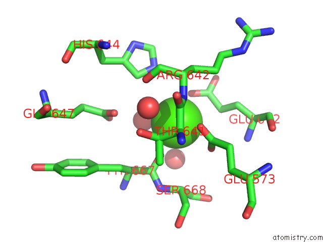
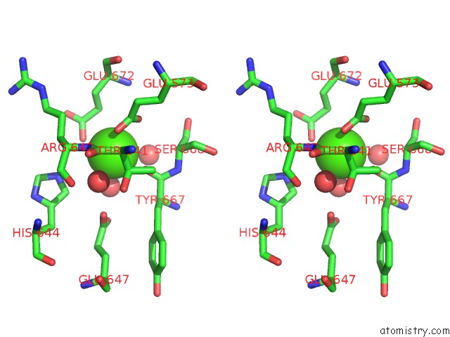
Calcium binding site 4 out of 4 in 1jrq
Go back to
Calcium binding site 4 out
of 4 in the X-Ray Structure Analysis of the Role of the Conserved Tyrosine-369 in Active Site of E. Coli Amine Oxidase
Mono view
Stereo pair view
Mono view
Stereo pair view
A full contact list of Calcium with other atoms in the Ca binding
site number 4 of X-Ray Structure Analysis of the Role of the Conserved Tyrosine-369 in Active Site of E. Coli Amine Oxidase within 5.0Å range:
|
Reference:
J.M.Murray,
C.R.Kurtis,
W.Tambyrajah,
C.G.Saysell,
C.M.Wilmot,
M.R.Parsons,
S.E.Phillips,
P.F.Knowles,
M.J.Mcpherson.
Conserved Tyrosine-369 in the Active Site of Escherichia Coli Copper Amine Oxidase Is Not Essential. Biochemistry V. 40 12808 2001.
ISSN: ISSN 0006-2960
PubMed: 11669617
DOI: 10.1021/BI011187P
Page generated: Mon Jul 7 16:13:24 2025
ISSN: ISSN 0006-2960
PubMed: 11669617
DOI: 10.1021/BI011187P
Last articles
Fe in 2YXOFe in 2YRS
Fe in 2YXC
Fe in 2YNM
Fe in 2YVJ
Fe in 2YP1
Fe in 2YU2
Fe in 2YU1
Fe in 2YQB
Fe in 2YOO