Calcium »
PDB 1jw6-1k9k »
1k1x »
Calcium in PDB 1k1x: Crystal Structure of 4-Alpha-Glucanotransferase From Thermococcus Litoralis
Enzymatic activity of Crystal Structure of 4-Alpha-Glucanotransferase From Thermococcus Litoralis
All present enzymatic activity of Crystal Structure of 4-Alpha-Glucanotransferase From Thermococcus Litoralis:
2.4.1.25;
2.4.1.25;
Protein crystallography data
The structure of Crystal Structure of 4-Alpha-Glucanotransferase From Thermococcus Litoralis, PDB code: 1k1x
was solved by
H.Imamura,
S.Fushinobu,
T.Kumasaka,
M.Yamamoto,
B.S.Jeon,
T.Wakagi,
H.Matsuzawa,
with X-Ray Crystallography technique. A brief refinement statistics is given in the table below:
| Resolution Low / High (Å) | 29.00 / 2.40 |
| Space group | P 21 21 2 |
| Cell size a, b, c (Å), α, β, γ (°) | 137.682, 161.494, 70.384, 90.00, 90.00, 90.00 |
| R / Rfree (%) | 19.5 / 23.6 |
Calcium Binding Sites:
The binding sites of Calcium atom in the Crystal Structure of 4-Alpha-Glucanotransferase From Thermococcus Litoralis
(pdb code 1k1x). This binding sites where shown within
5.0 Angstroms radius around Calcium atom.
In total 3 binding sites of Calcium where determined in the Crystal Structure of 4-Alpha-Glucanotransferase From Thermococcus Litoralis, PDB code: 1k1x:
Jump to Calcium binding site number: 1; 2; 3;
In total 3 binding sites of Calcium where determined in the Crystal Structure of 4-Alpha-Glucanotransferase From Thermococcus Litoralis, PDB code: 1k1x:
Jump to Calcium binding site number: 1; 2; 3;
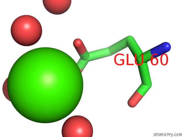
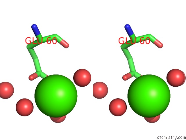
Calcium binding site 1 out of 3 in 1k1x
Go back to
Calcium binding site 1 out
of 3 in the Crystal Structure of 4-Alpha-Glucanotransferase From Thermococcus Litoralis
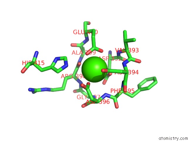
Mono view
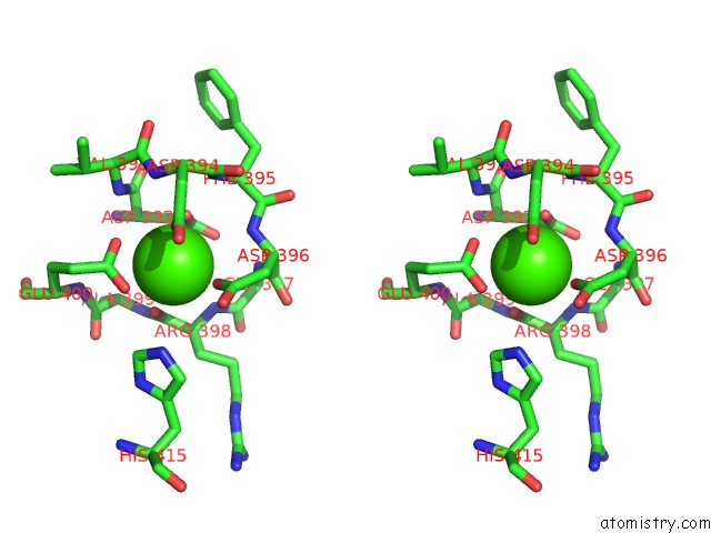
Stereo pair view
Mono view
Stereo pair view
A full contact list of Calcium with other atoms in the Ca binding
site number 1 of Crystal Structure of 4-Alpha-Glucanotransferase From Thermococcus Litoralis within 5.0Å range:
|
Calcium binding site 2 out of 3 in 1k1x
Go back to
Calcium binding site 2 out
of 3 in the Crystal Structure of 4-Alpha-Glucanotransferase From Thermococcus Litoralis
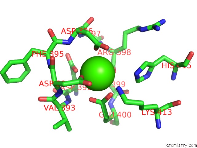
Mono view
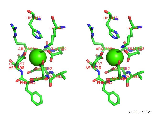
Stereo pair view
Mono view
Stereo pair view
A full contact list of Calcium with other atoms in the Ca binding
site number 2 of Crystal Structure of 4-Alpha-Glucanotransferase From Thermococcus Litoralis within 5.0Å range:
|
Calcium binding site 3 out of 3 in 1k1x
Go back to
Calcium binding site 3 out
of 3 in the Crystal Structure of 4-Alpha-Glucanotransferase From Thermococcus Litoralis
Mono view
Stereo pair view
Mono view
Stereo pair view
A full contact list of Calcium with other atoms in the Ca binding
site number 3 of Crystal Structure of 4-Alpha-Glucanotransferase From Thermococcus Litoralis within 5.0Å range:
|
Reference:
H.Imamura,
S.Fushinobu,
M.Yamamoto,
T.Kumasaka,
B.S.Jeon,
T.Wakagi,
H.Matsuzawa.
Crystal Structures of 4-Alpha-Glucanotransferase From Thermococcus Litoralis and Its Complex with An Inhibitor J.Biol.Chem. V. 278 19378 2003.
ISSN: ISSN 0021-9258
PubMed: 12618437
DOI: 10.1074/JBC.M213134200
Page generated: Thu Jul 11 11:08:18 2024
ISSN: ISSN 0021-9258
PubMed: 12618437
DOI: 10.1074/JBC.M213134200
Last articles
Zn in 9MJ5Zn in 9HNW
Zn in 9G0L
Zn in 9FNE
Zn in 9DZN
Zn in 9E0I
Zn in 9D32
Zn in 9DAK
Zn in 8ZXC
Zn in 8ZUF