Calcium »
PDB 1jw6-1k9k »
1k72 »
Calcium in PDB 1k72: The X-Ray Crystal Structure of CEL9G Complexed with Cellotriose
Enzymatic activity of The X-Ray Crystal Structure of CEL9G Complexed with Cellotriose
All present enzymatic activity of The X-Ray Crystal Structure of CEL9G Complexed with Cellotriose:
3.2.1.4;
3.2.1.4;
Protein crystallography data
The structure of The X-Ray Crystal Structure of CEL9G Complexed with Cellotriose, PDB code: 1k72
was solved by
D.Mandelman,
A.Belaich,
J.P.Belaich,
N.Aghajari,
H.Driguez,
R.Haser,
with X-Ray Crystallography technique. A brief refinement statistics is given in the table below:
| Resolution Low / High (Å) | 36.31 / 1.80 |
| Space group | P 1 |
| Cell size a, b, c (Å), α, β, γ (°) | 57.000, 57.600, 86.800, 93.60, 100.80, 99.50 |
| R / Rfree (%) | 16.6 / 19.8 |
Other elements in 1k72:
The structure of The X-Ray Crystal Structure of CEL9G Complexed with Cellotriose also contains other interesting chemical elements:
| Magnesium | (Mg) | 4 atoms |
Calcium Binding Sites:
The binding sites of Calcium atom in the The X-Ray Crystal Structure of CEL9G Complexed with Cellotriose
(pdb code 1k72). This binding sites where shown within
5.0 Angstroms radius around Calcium atom.
In total 4 binding sites of Calcium where determined in the The X-Ray Crystal Structure of CEL9G Complexed with Cellotriose, PDB code: 1k72:
Jump to Calcium binding site number: 1; 2; 3; 4;
In total 4 binding sites of Calcium where determined in the The X-Ray Crystal Structure of CEL9G Complexed with Cellotriose, PDB code: 1k72:
Jump to Calcium binding site number: 1; 2; 3; 4;
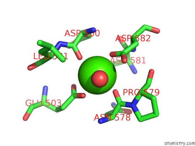
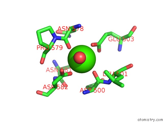
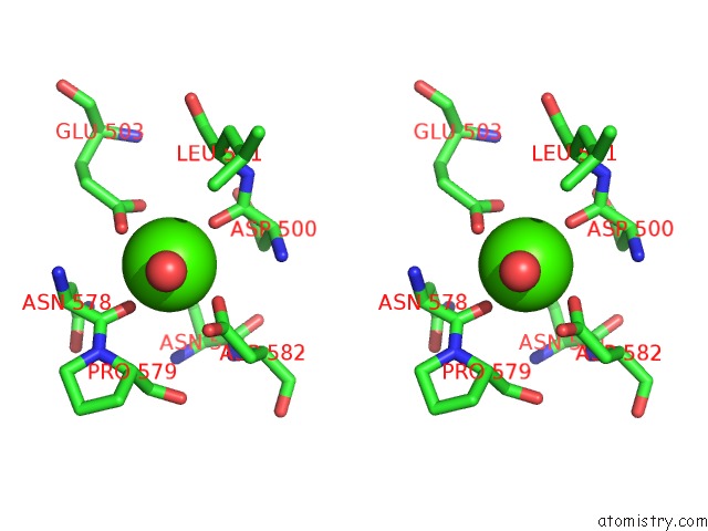
Calcium binding site 1 out of 4 in 1k72
Go back to
Calcium binding site 1 out
of 4 in the The X-Ray Crystal Structure of CEL9G Complexed with Cellotriose
Mono view
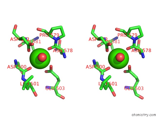
Stereo pair view
Mono view
Stereo pair view
A full contact list of Calcium with other atoms in the Ca binding
site number 1 of The X-Ray Crystal Structure of CEL9G Complexed with Cellotriose within 5.0Å range:
|
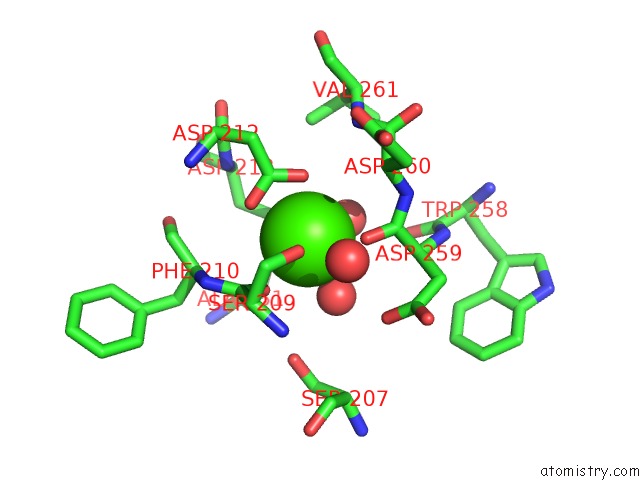
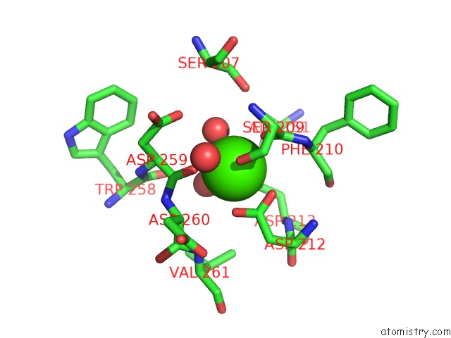
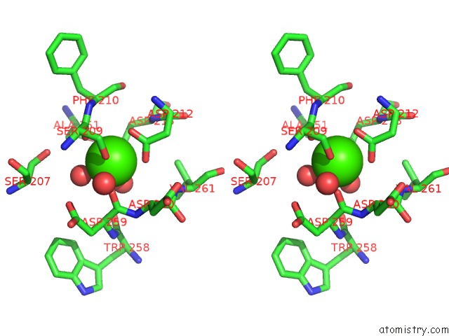
Calcium binding site 2 out of 4 in 1k72
Go back to
Calcium binding site 2 out
of 4 in the The X-Ray Crystal Structure of CEL9G Complexed with Cellotriose
Mono view
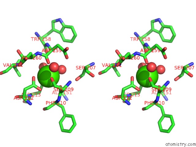
Stereo pair view
Mono view
Stereo pair view
A full contact list of Calcium with other atoms in the Ca binding
site number 2 of The X-Ray Crystal Structure of CEL9G Complexed with Cellotriose within 5.0Å range:
|
Calcium binding site 3 out of 4 in 1k72
Go back to
Calcium binding site 3 out
of 4 in the The X-Ray Crystal Structure of CEL9G Complexed with Cellotriose
Mono view
Stereo pair view
Mono view
Stereo pair view
A full contact list of Calcium with other atoms in the Ca binding
site number 3 of The X-Ray Crystal Structure of CEL9G Complexed with Cellotriose within 5.0Å range:
|
Calcium binding site 4 out of 4 in 1k72
Go back to
Calcium binding site 4 out
of 4 in the The X-Ray Crystal Structure of CEL9G Complexed with Cellotriose
Mono view
Stereo pair view
Mono view
Stereo pair view
A full contact list of Calcium with other atoms in the Ca binding
site number 4 of The X-Ray Crystal Structure of CEL9G Complexed with Cellotriose within 5.0Å range:
|
Reference:
D.Mandelman,
A.Belaich,
J.P.Belaich,
N.Aghajari,
H.Driguez,
R.Haser.
X-Ray Crystal Structure of the Multidomain Endoglucanase CEL9G From Clostridium Cellulolyticum Complexed with Natural and Synthetic Cello-Oligosaccharides J.Bacteriol. V. 185 4127 2003.
ISSN: ISSN 0021-9193
PubMed: 12837787
DOI: 10.1128/JB.185.14.4127-4135.2003
Page generated: Mon Jul 7 16:19:17 2025
ISSN: ISSN 0021-9193
PubMed: 12837787
DOI: 10.1128/JB.185.14.4127-4135.2003
Last articles
F in 4ISFF in 4IKS
F in 4ISE
F in 4IQV
F in 4IQW
F in 4IQT
F in 4IQU
F in 4INB
F in 4IKT
F in 4IJU