Calcium »
PDB 1jw6-1k9k »
1k90 »
Calcium in PDB 1k90: Crystal Structure of the Adenylyl Cyclase Domain of Anthrax Edema Factor (Ef) in Complex with Calmodulin and 3' Deoxy-Atp
Enzymatic activity of Crystal Structure of the Adenylyl Cyclase Domain of Anthrax Edema Factor (Ef) in Complex with Calmodulin and 3' Deoxy-Atp
All present enzymatic activity of Crystal Structure of the Adenylyl Cyclase Domain of Anthrax Edema Factor (Ef) in Complex with Calmodulin and 3' Deoxy-Atp:
4.6.1.1;
4.6.1.1;
Protein crystallography data
The structure of Crystal Structure of the Adenylyl Cyclase Domain of Anthrax Edema Factor (Ef) in Complex with Calmodulin and 3' Deoxy-Atp, PDB code: 1k90
was solved by
C.L.Drum,
S.-Z.Yan,
J.Bard,
Y.-Q.Shen,
D.Lu,
S.Soelaiman,
Z.Grabarek,
A.Bohm,
W.-J.Tang,
with X-Ray Crystallography technique. A brief refinement statistics is given in the table below:
| Resolution Low / High (Å) | 19.97 / 2.75 |
| Space group | I 2 2 2 |
| Cell size a, b, c (Å), α, β, γ (°) | 116.099, 166.100, 343.330, 90.00, 90.00, 90.00 |
| R / Rfree (%) | 21.5 / 28.6 |
Other elements in 1k90:
The structure of Crystal Structure of the Adenylyl Cyclase Domain of Anthrax Edema Factor (Ef) in Complex with Calmodulin and 3' Deoxy-Atp also contains other interesting chemical elements:
| Ytterbium | (Yb) | 3 atoms |
Calcium Binding Sites:
The binding sites of Calcium atom in the Crystal Structure of the Adenylyl Cyclase Domain of Anthrax Edema Factor (Ef) in Complex with Calmodulin and 3' Deoxy-Atp
(pdb code 1k90). This binding sites where shown within
5.0 Angstroms radius around Calcium atom.
In total 6 binding sites of Calcium where determined in the Crystal Structure of the Adenylyl Cyclase Domain of Anthrax Edema Factor (Ef) in Complex with Calmodulin and 3' Deoxy-Atp, PDB code: 1k90:
Jump to Calcium binding site number: 1; 2; 3; 4; 5; 6;
In total 6 binding sites of Calcium where determined in the Crystal Structure of the Adenylyl Cyclase Domain of Anthrax Edema Factor (Ef) in Complex with Calmodulin and 3' Deoxy-Atp, PDB code: 1k90:
Jump to Calcium binding site number: 1; 2; 3; 4; 5; 6;
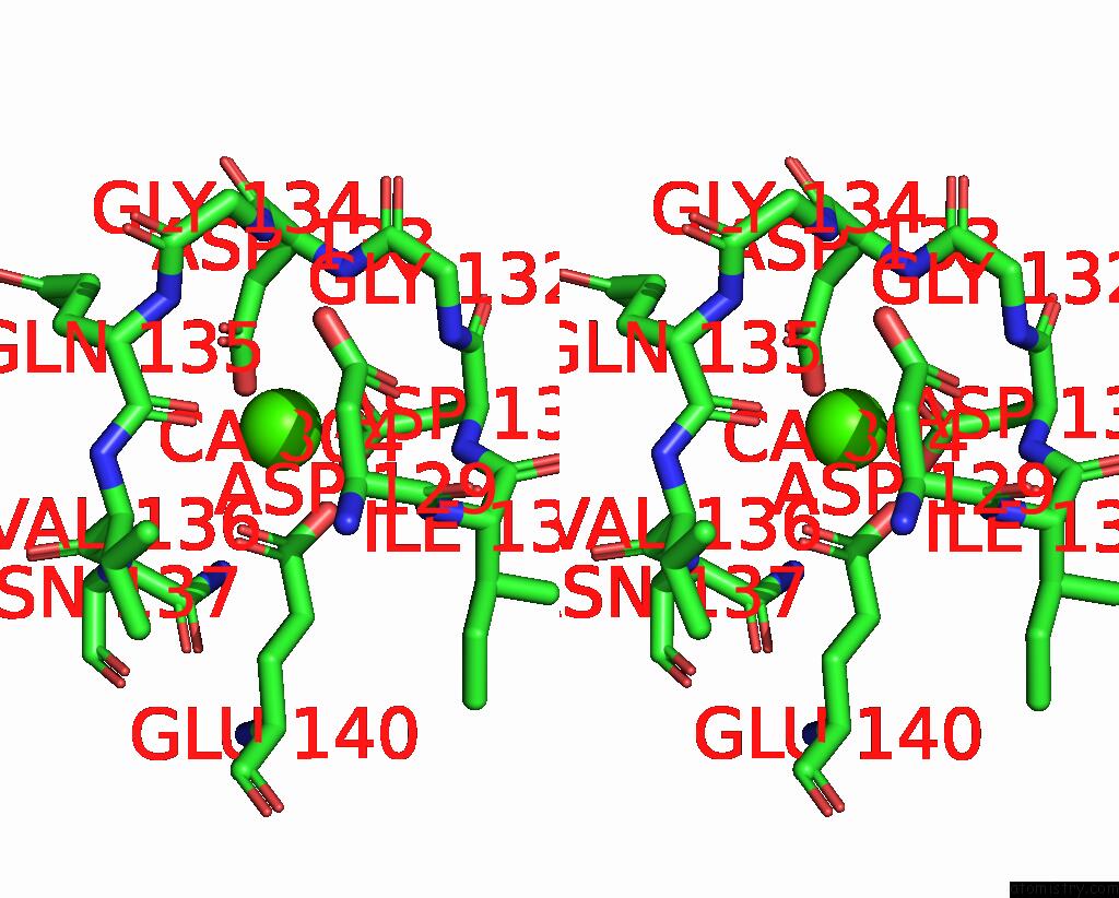
Calcium binding site 1 out of 6 in 1k90
Go back to
Calcium binding site 1 out
of 6 in the Crystal Structure of the Adenylyl Cyclase Domain of Anthrax Edema Factor (Ef) in Complex with Calmodulin and 3' Deoxy-Atp

Mono view

Stereo pair view
Mono view
Stereo pair view
A full contact list of Calcium with other atoms in the Ca binding
site number 1 of Crystal Structure of the Adenylyl Cyclase Domain of Anthrax Edema Factor (Ef) in Complex with Calmodulin and 3' Deoxy-Atp within 5.0Å range:
|
Calcium binding site 2 out of 6 in 1k90
Go back to
Calcium binding site 2 out
of 6 in the Crystal Structure of the Adenylyl Cyclase Domain of Anthrax Edema Factor (Ef) in Complex with Calmodulin and 3' Deoxy-Atp

Mono view

Stereo pair view
Mono view
Stereo pair view
A full contact list of Calcium with other atoms in the Ca binding
site number 2 of Crystal Structure of the Adenylyl Cyclase Domain of Anthrax Edema Factor (Ef) in Complex with Calmodulin and 3' Deoxy-Atp within 5.0Å range:
|
Calcium binding site 3 out of 6 in 1k90
Go back to
Calcium binding site 3 out
of 6 in the Crystal Structure of the Adenylyl Cyclase Domain of Anthrax Edema Factor (Ef) in Complex with Calmodulin and 3' Deoxy-Atp

Mono view

Stereo pair view
Mono view
Stereo pair view
A full contact list of Calcium with other atoms in the Ca binding
site number 3 of Crystal Structure of the Adenylyl Cyclase Domain of Anthrax Edema Factor (Ef) in Complex with Calmodulin and 3' Deoxy-Atp within 5.0Å range:
|
Calcium binding site 4 out of 6 in 1k90
Go back to
Calcium binding site 4 out
of 6 in the Crystal Structure of the Adenylyl Cyclase Domain of Anthrax Edema Factor (Ef) in Complex with Calmodulin and 3' Deoxy-Atp

Mono view

Stereo pair view
Mono view
Stereo pair view
A full contact list of Calcium with other atoms in the Ca binding
site number 4 of Crystal Structure of the Adenylyl Cyclase Domain of Anthrax Edema Factor (Ef) in Complex with Calmodulin and 3' Deoxy-Atp within 5.0Å range:
|
Calcium binding site 5 out of 6 in 1k90
Go back to
Calcium binding site 5 out
of 6 in the Crystal Structure of the Adenylyl Cyclase Domain of Anthrax Edema Factor (Ef) in Complex with Calmodulin and 3' Deoxy-Atp

Mono view
Stereo pair view
Mono view
Stereo pair view
A full contact list of Calcium with other atoms in the Ca binding
site number 5 of Crystal Structure of the Adenylyl Cyclase Domain of Anthrax Edema Factor (Ef) in Complex with Calmodulin and 3' Deoxy-Atp within 5.0Å range:
|
Calcium binding site 6 out of 6 in 1k90
Go back to
Calcium binding site 6 out
of 6 in the Crystal Structure of the Adenylyl Cyclase Domain of Anthrax Edema Factor (Ef) in Complex with Calmodulin and 3' Deoxy-Atp

Mono view

Stereo pair view
Mono view
Stereo pair view
A full contact list of Calcium with other atoms in the Ca binding
site number 6 of Crystal Structure of the Adenylyl Cyclase Domain of Anthrax Edema Factor (Ef) in Complex with Calmodulin and 3' Deoxy-Atp within 5.0Å range:
|
Reference:
C.L.Drum,
S.-Z.Yan,
J.Bard,
Y.-Q.Shen,
D.Lu,
S.Soelaiman,
Z.Grabarek,
A.Bohm,
W.-J.Tang.
Structural Basis For the Activation of Anthrax Adenylyl Cyclase Exotoxin By Calmodulin. Nature V. 415 396 2002.
ISSN: ISSN 0028-0836
PubMed: 11807546
DOI: 10.1038/415396A
Page generated: Mon Jul 7 16:20:26 2025
ISSN: ISSN 0028-0836
PubMed: 11807546
DOI: 10.1038/415396A
Last articles
Cl in 8CW3Cl in 8CW1
Cl in 8CVW
Cl in 8CW0
Cl in 8CVV
Cl in 8CVU
Cl in 8CUR
Cl in 8CU9
Cl in 8CTY
Cl in 8CTM