Calcium »
PDB 1jw6-1k9k »
1k96 »
Calcium in PDB 1k96: Crystal Structure of Calcium Bound Human S100A6
Protein crystallography data
The structure of Crystal Structure of Calcium Bound Human S100A6, PDB code: 1k96
was solved by
L.R.Otterbein,
R.Dominguez,
with X-Ray Crystallography technique. A brief refinement statistics is given in the table below:
| Resolution Low / High (Å) | 15.00 / 1.44 |
| Space group | C 1 2 1 |
| Cell size a, b, c (Å), α, β, γ (°) | 45.180, 39.230, 48.110, 90.00, 110.44, 90.00 |
| R / Rfree (%) | 21.4 / 22.6 |
Calcium Binding Sites:
The binding sites of Calcium atom in the Crystal Structure of Calcium Bound Human S100A6
(pdb code 1k96). This binding sites where shown within
5.0 Angstroms radius around Calcium atom.
In total 2 binding sites of Calcium where determined in the Crystal Structure of Calcium Bound Human S100A6, PDB code: 1k96:
Jump to Calcium binding site number: 1; 2;
In total 2 binding sites of Calcium where determined in the Crystal Structure of Calcium Bound Human S100A6, PDB code: 1k96:
Jump to Calcium binding site number: 1; 2;
Calcium binding site 1 out of 2 in 1k96
Go back to
Calcium binding site 1 out
of 2 in the Crystal Structure of Calcium Bound Human S100A6
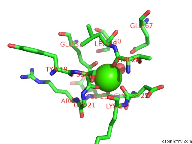
Mono view
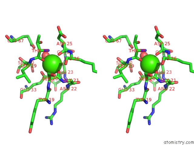
Stereo pair view
Mono view
Stereo pair view
A full contact list of Calcium with other atoms in the Ca binding
site number 1 of Crystal Structure of Calcium Bound Human S100A6 within 5.0Å range:
|
Calcium binding site 2 out of 2 in 1k96
Go back to
Calcium binding site 2 out
of 2 in the Crystal Structure of Calcium Bound Human S100A6
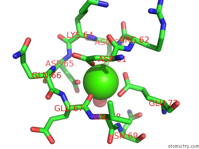
Mono view
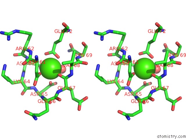
Stereo pair view
Mono view
Stereo pair view
A full contact list of Calcium with other atoms in the Ca binding
site number 2 of Crystal Structure of Calcium Bound Human S100A6 within 5.0Å range:
|
Reference:
L.R.Otterbein,
J.Kordowska,
C.Witte-Hoffmann,
C.L.Wang,
R.Dominguez.
Crystal Structures of S100A6 in the Ca(2+)-Free and Ca(2+)-Bound States: the Calcium Sensor Mechanism of S100 Proteins Revealed at Atomic Resolution. Structure V. 10 557 2002.
ISSN: ISSN 0969-2126
PubMed: 11937060
DOI: 10.1016/S0969-2126(02)00740-2
Page generated: Mon Jul 7 16:22:25 2025
ISSN: ISSN 0969-2126
PubMed: 11937060
DOI: 10.1016/S0969-2126(02)00740-2
Last articles
Ca in 1QAFCa in 1QA0
Ca in 1Q9Y
Ca in 1Q9X
Ca in 1Q8V
Ca in 1Q8S
Ca in 1Q8Q
Ca in 1Q8P
Ca in 1Q8O
Ca in 1Q8H