Calcium »
PDB 1k9u-1kre »
1kdq »
Calcium in PDB 1kdq: Crystal Structure Analysis of the Mutant S189D Rat Chymotrypsin
Enzymatic activity of Crystal Structure Analysis of the Mutant S189D Rat Chymotrypsin
All present enzymatic activity of Crystal Structure Analysis of the Mutant S189D Rat Chymotrypsin:
3.4.21.1;
3.4.21.1;
Protein crystallography data
The structure of Crystal Structure Analysis of the Mutant S189D Rat Chymotrypsin, PDB code: 1kdq
was solved by
E.Szabo,
Z.Bocskei,
G.Naray-Szabo,
L.Graf,
I.Venekei,
with X-Ray Crystallography technique. A brief refinement statistics is given in the table below:
| Resolution Low / High (Å) | 32.97 / 2.55 |
| Space group | C 2 2 21 |
| Cell size a, b, c (Å), α, β, γ (°) | 74.589, 76.707, 83.478, 90.00, 90.00, 90.00 |
| R / Rfree (%) | 22.1 / 27.7 |
Calcium Binding Sites:
The binding sites of Calcium atom in the Crystal Structure Analysis of the Mutant S189D Rat Chymotrypsin
(pdb code 1kdq). This binding sites where shown within
5.0 Angstroms radius around Calcium atom.
In total only one binding site of Calcium was determined in the Crystal Structure Analysis of the Mutant S189D Rat Chymotrypsin, PDB code: 1kdq:
In total only one binding site of Calcium was determined in the Crystal Structure Analysis of the Mutant S189D Rat Chymotrypsin, PDB code: 1kdq:
Calcium binding site 1 out of 1 in 1kdq
Go back to
Calcium binding site 1 out
of 1 in the Crystal Structure Analysis of the Mutant S189D Rat Chymotrypsin
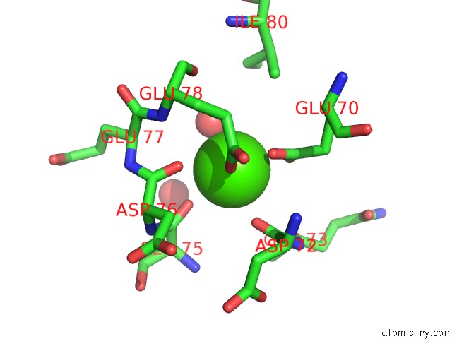
Mono view
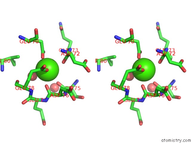
Stereo pair view
Mono view
Stereo pair view
A full contact list of Calcium with other atoms in the Ca binding
site number 1 of Crystal Structure Analysis of the Mutant S189D Rat Chymotrypsin within 5.0Å range:
|
Reference:
E.Szabo,
I.Venekei,
Z.Bocskei,
G.Naray-Szabo,
L.Graf.
Three Dimensional Structures of S189D Chymotrypsin and D189S Trypsin Mutants: the Effect of Polarity at Site 189 on A Protease-Specific Stabilization of the Substrate-Binding Site J.Mol.Biol. V. 331 1121 2003.
ISSN: ISSN 0022-2836
PubMed: 12927546
DOI: 10.1016/S0022-2836(03)00849-0
Page generated: Thu Jul 11 11:20:22 2024
ISSN: ISSN 0022-2836
PubMed: 12927546
DOI: 10.1016/S0022-2836(03)00849-0
Last articles
Zn in 9J0NZn in 9J0O
Zn in 9J0P
Zn in 9FJX
Zn in 9EKB
Zn in 9C0F
Zn in 9CAH
Zn in 9CH0
Zn in 9CH3
Zn in 9CH1