Calcium »
PDB 1krf-1l3f »
1krf »
Calcium in PDB 1krf: Structure of P. Citrinum Alpha 1,2-Mannosidase Reveals the Basis For Differences in Specificity of the Er and Golgi Class I Enzymes
Enzymatic activity of Structure of P. Citrinum Alpha 1,2-Mannosidase Reveals the Basis For Differences in Specificity of the Er and Golgi Class I Enzymes
All present enzymatic activity of Structure of P. Citrinum Alpha 1,2-Mannosidase Reveals the Basis For Differences in Specificity of the Er and Golgi Class I Enzymes:
3.2.1.113;
3.2.1.113;
Protein crystallography data
The structure of Structure of P. Citrinum Alpha 1,2-Mannosidase Reveals the Basis For Differences in Specificity of the Er and Golgi Class I Enzymes, PDB code: 1krf
was solved by
Y.D.Lobsanov,
F.Vallee,
A.Imberty,
T.Yoshida,
P.Yip,
A.Herscovics,
P.L.Howell,
with X-Ray Crystallography technique. A brief refinement statistics is given in the table below:
| Resolution Low / High (Å) | 50.00 / 2.20 |
| Space group | P 1 21 1 |
| Cell size a, b, c (Å), α, β, γ (°) | 56.487, 110.997, 86.235, 90.00, 99.17, 90.00 |
| R / Rfree (%) | 19.9 / 23.6 |
Calcium Binding Sites:
The binding sites of Calcium atom in the Structure of P. Citrinum Alpha 1,2-Mannosidase Reveals the Basis For Differences in Specificity of the Er and Golgi Class I Enzymes
(pdb code 1krf). This binding sites where shown within
5.0 Angstroms radius around Calcium atom.
In total 2 binding sites of Calcium where determined in the Structure of P. Citrinum Alpha 1,2-Mannosidase Reveals the Basis For Differences in Specificity of the Er and Golgi Class I Enzymes, PDB code: 1krf:
Jump to Calcium binding site number: 1; 2;
In total 2 binding sites of Calcium where determined in the Structure of P. Citrinum Alpha 1,2-Mannosidase Reveals the Basis For Differences in Specificity of the Er and Golgi Class I Enzymes, PDB code: 1krf:
Jump to Calcium binding site number: 1; 2;
Calcium binding site 1 out of 2 in 1krf
Go back to
Calcium binding site 1 out
of 2 in the Structure of P. Citrinum Alpha 1,2-Mannosidase Reveals the Basis For Differences in Specificity of the Er and Golgi Class I Enzymes
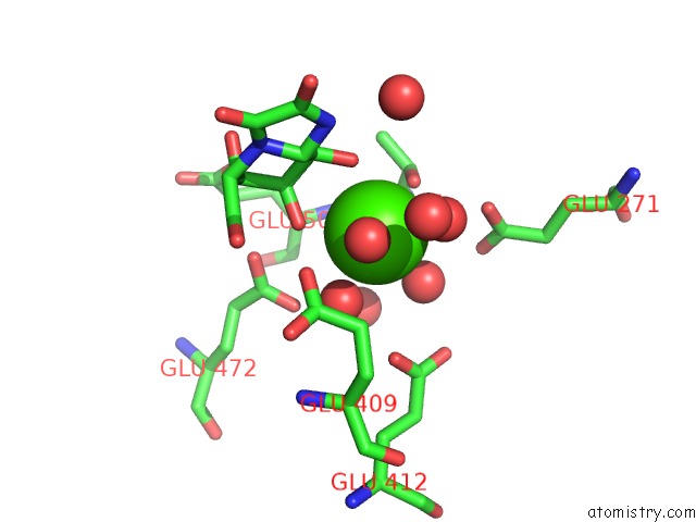
Mono view
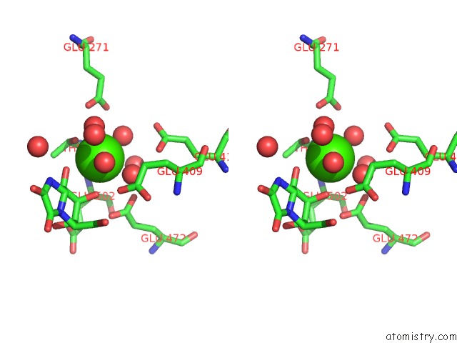
Stereo pair view
Mono view
Stereo pair view
A full contact list of Calcium with other atoms in the Ca binding
site number 1 of Structure of P. Citrinum Alpha 1,2-Mannosidase Reveals the Basis For Differences in Specificity of the Er and Golgi Class I Enzymes within 5.0Å range:
|
Calcium binding site 2 out of 2 in 1krf
Go back to
Calcium binding site 2 out
of 2 in the Structure of P. Citrinum Alpha 1,2-Mannosidase Reveals the Basis For Differences in Specificity of the Er and Golgi Class I Enzymes
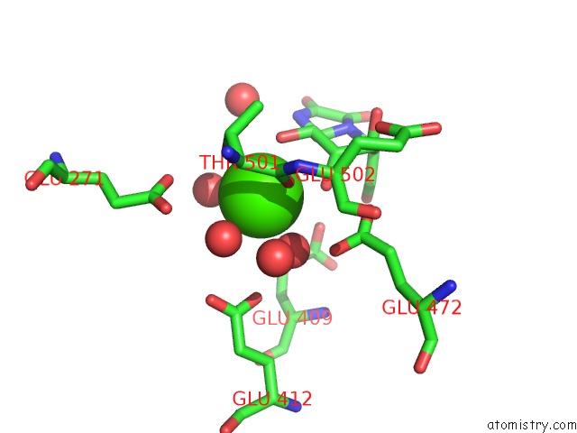
Mono view
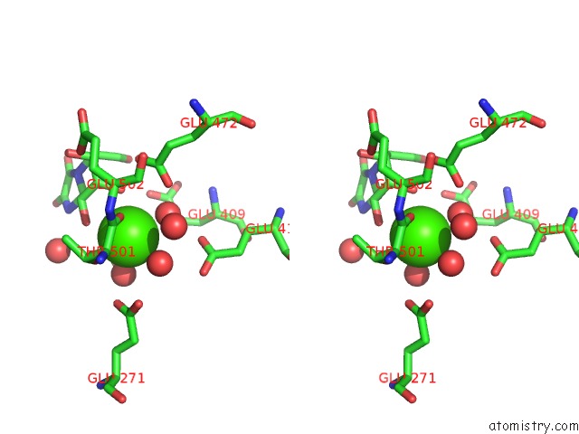
Stereo pair view
Mono view
Stereo pair view
A full contact list of Calcium with other atoms in the Ca binding
site number 2 of Structure of P. Citrinum Alpha 1,2-Mannosidase Reveals the Basis For Differences in Specificity of the Er and Golgi Class I Enzymes within 5.0Å range:
|
Reference:
Y.D.Lobsanov,
F.Vallee,
A.Imberty,
T.Yoshida,
P.Yip,
A.Herscovics,
P.L.Howell.
Structure of Penicillium Citrinum Alpha 1,2-Mannosidase Reveals the Basis For Differences in Specificity of the Endoplasmic Reticulum and Golgi Class I Enzymes. J.Biol.Chem. V. 277 5620 2002.
ISSN: ISSN 0021-9258
PubMed: 11714724
DOI: 10.1074/JBC.M110243200
Page generated: Thu Jul 11 11:29:02 2024
ISSN: ISSN 0021-9258
PubMed: 11714724
DOI: 10.1074/JBC.M110243200
Last articles
Zn in 9MJ5Zn in 9HNW
Zn in 9G0L
Zn in 9FNE
Zn in 9DZN
Zn in 9E0I
Zn in 9D32
Zn in 9DAK
Zn in 8ZXC
Zn in 8ZUF