Calcium »
PDB 1krf-1l3f »
1kvw »
Calcium in PDB 1kvw: Carboxylic Ester Hydrolase, Single Mutant H48Q of Bovine Pancreatic PLA2 Enzyme
Enzymatic activity of Carboxylic Ester Hydrolase, Single Mutant H48Q of Bovine Pancreatic PLA2 Enzyme
All present enzymatic activity of Carboxylic Ester Hydrolase, Single Mutant H48Q of Bovine Pancreatic PLA2 Enzyme:
3.1.1.4;
3.1.1.4;
Protein crystallography data
The structure of Carboxylic Ester Hydrolase, Single Mutant H48Q of Bovine Pancreatic PLA2 Enzyme, PDB code: 1kvw
was solved by
M.Sundaralingam,
with X-Ray Crystallography technique. A brief refinement statistics is given in the table below:
| Resolution Low / High (Å) | 10.00 / 1.95 |
| Space group | P 31 2 1 |
| Cell size a, b, c (Å), α, β, γ (°) | 47.120, 47.120, 102.880, 90.00, 90.00, 120.00 |
| R / Rfree (%) | 20.9 / 31.4 |
Calcium Binding Sites:
The binding sites of Calcium atom in the Carboxylic Ester Hydrolase, Single Mutant H48Q of Bovine Pancreatic PLA2 Enzyme
(pdb code 1kvw). This binding sites where shown within
5.0 Angstroms radius around Calcium atom.
In total only one binding site of Calcium was determined in the Carboxylic Ester Hydrolase, Single Mutant H48Q of Bovine Pancreatic PLA2 Enzyme, PDB code: 1kvw:
In total only one binding site of Calcium was determined in the Carboxylic Ester Hydrolase, Single Mutant H48Q of Bovine Pancreatic PLA2 Enzyme, PDB code: 1kvw:
Calcium binding site 1 out of 1 in 1kvw
Go back to
Calcium binding site 1 out
of 1 in the Carboxylic Ester Hydrolase, Single Mutant H48Q of Bovine Pancreatic PLA2 Enzyme
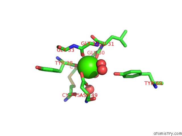
Mono view
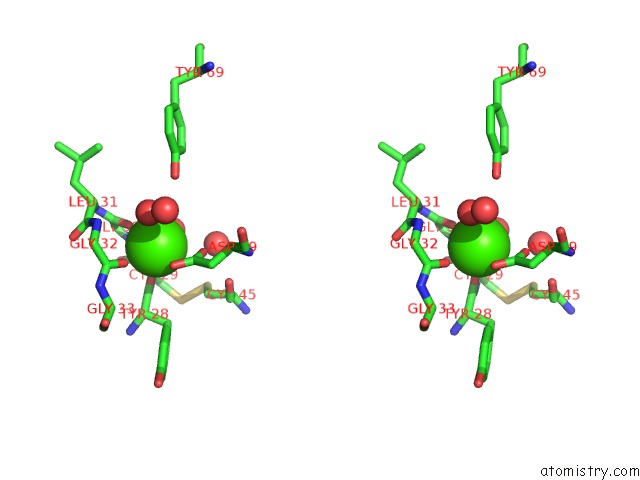
Stereo pair view
Mono view
Stereo pair view
A full contact list of Calcium with other atoms in the Ca binding
site number 1 of Carboxylic Ester Hydrolase, Single Mutant H48Q of Bovine Pancreatic PLA2 Enzyme within 5.0Å range:
|
Reference:
K.Sekar,
R.Biswas,
Y.Li,
M.Tsai,
M.Sundaralingam.
Structures of the Catalytic Site Mutants D99A and H48Q and the Calcium-Loop Mutant D49E of Phospholipase A2. Acta Crystallogr.,Sect.D V. 55 443 1999.
ISSN: ISSN 0907-4449
PubMed: 10089353
DOI: 10.1107/S0907444998013699
Page generated: Thu Jul 11 11:30:27 2024
ISSN: ISSN 0907-4449
PubMed: 10089353
DOI: 10.1107/S0907444998013699
Last articles
Zn in 9MJ5Zn in 9HNW
Zn in 9G0L
Zn in 9FNE
Zn in 9DZN
Zn in 9E0I
Zn in 9D32
Zn in 9DAK
Zn in 8ZXC
Zn in 8ZUF