Calcium »
PDB 1krf-1l3f »
1kxh »
Calcium in PDB 1kxh: Crystal Structure of the Complex Between An Inactive Mutant of Psychrophilic Alpha-Amylase (D174N) and Acarbose
Enzymatic activity of Crystal Structure of the Complex Between An Inactive Mutant of Psychrophilic Alpha-Amylase (D174N) and Acarbose
All present enzymatic activity of Crystal Structure of the Complex Between An Inactive Mutant of Psychrophilic Alpha-Amylase (D174N) and Acarbose:
3.2.1.1;
3.2.1.1;
Protein crystallography data
The structure of Crystal Structure of the Complex Between An Inactive Mutant of Psychrophilic Alpha-Amylase (D174N) and Acarbose, PDB code: 1kxh
was solved by
N.Aghajari,
R.Haser,
with X-Ray Crystallography technique. A brief refinement statistics is given in the table below:
| Resolution Low / High (Å) | 27.70 / 2.30 |
| Space group | C 2 2 21 |
| Cell size a, b, c (Å), α, β, γ (°) | 71.100, 139.900, 115.300, 90.00, 90.00, 90.00 |
| R / Rfree (%) | 13.8 / 18.8 |
Other elements in 1kxh:
The structure of Crystal Structure of the Complex Between An Inactive Mutant of Psychrophilic Alpha-Amylase (D174N) and Acarbose also contains other interesting chemical elements:
| Chlorine | (Cl) | 1 atom |
Calcium Binding Sites:
The binding sites of Calcium atom in the Crystal Structure of the Complex Between An Inactive Mutant of Psychrophilic Alpha-Amylase (D174N) and Acarbose
(pdb code 1kxh). This binding sites where shown within
5.0 Angstroms radius around Calcium atom.
In total only one binding site of Calcium was determined in the Crystal Structure of the Complex Between An Inactive Mutant of Psychrophilic Alpha-Amylase (D174N) and Acarbose, PDB code: 1kxh:
In total only one binding site of Calcium was determined in the Crystal Structure of the Complex Between An Inactive Mutant of Psychrophilic Alpha-Amylase (D174N) and Acarbose, PDB code: 1kxh:
Calcium binding site 1 out of 1 in 1kxh
Go back to
Calcium binding site 1 out
of 1 in the Crystal Structure of the Complex Between An Inactive Mutant of Psychrophilic Alpha-Amylase (D174N) and Acarbose
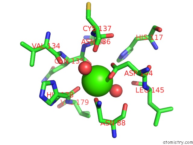
Mono view
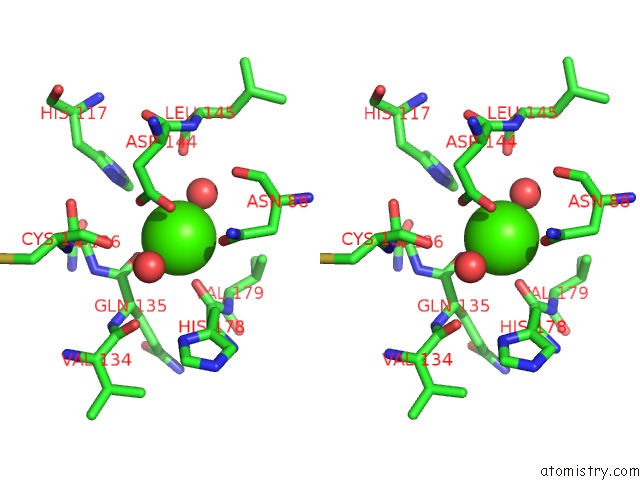
Stereo pair view
Mono view
Stereo pair view
A full contact list of Calcium with other atoms in the Ca binding
site number 1 of Crystal Structure of the Complex Between An Inactive Mutant of Psychrophilic Alpha-Amylase (D174N) and Acarbose within 5.0Å range:
|
Reference:
N.Aghajari,
M.Roth,
R.Haser.
Crystallographic Evidence of A Transglycosylation Reaction: Ternary Complexes of A Psychrophilic Alpha-Amylase. Biochemistry V. 41 4273.
ISSN: ISSN 0006-2960
PubMed: 11914073
DOI: 10.1021/BI0160516
Page generated: Mon Jul 7 16:40:42 2025
ISSN: ISSN 0006-2960
PubMed: 11914073
DOI: 10.1021/BI0160516
Last articles
F in 4L6QF in 4L7F
F in 4L4L
F in 4L46
F in 4L45
F in 4L44
F in 4L43
F in 4L42
F in 4L3L
F in 4L3J