Calcium »
PDB 1krf-1l3f »
1l2o »
Calcium in PDB 1l2o: Scallop Myosin S1-Adp-P-Pdm in the Actin-Detached Conformation
Protein crystallography data
The structure of Scallop Myosin S1-Adp-P-Pdm in the Actin-Detached Conformation, PDB code: 1l2o
was solved by
M.Himmel,
S.Gourinath,
L.Reshetnikova,
Y.Shen,
G.Szent-Gyorgyi,
C.Cohen,
with X-Ray Crystallography technique. A brief refinement statistics is given in the table below:
| Resolution Low / High (Å) | 54.16 / 2.80 |
| Space group | P 1 |
| Cell size a, b, c (Å), α, β, γ (°) | 51.800, 57.000, 150.500, 95.60, 96.30, 101.50 |
| R / Rfree (%) | 28 / 32.7 |
Other elements in 1l2o:
The structure of Scallop Myosin S1-Adp-P-Pdm in the Actin-Detached Conformation also contains other interesting chemical elements:
| Magnesium | (Mg) | 2 atoms |
Calcium Binding Sites:
The binding sites of Calcium atom in the Scallop Myosin S1-Adp-P-Pdm in the Actin-Detached Conformation
(pdb code 1l2o). This binding sites where shown within
5.0 Angstroms radius around Calcium atom.
In total only one binding site of Calcium was determined in the Scallop Myosin S1-Adp-P-Pdm in the Actin-Detached Conformation, PDB code: 1l2o:
In total only one binding site of Calcium was determined in the Scallop Myosin S1-Adp-P-Pdm in the Actin-Detached Conformation, PDB code: 1l2o:
Calcium binding site 1 out of 1 in 1l2o
Go back to
Calcium binding site 1 out
of 1 in the Scallop Myosin S1-Adp-P-Pdm in the Actin-Detached Conformation
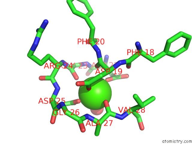
Mono view
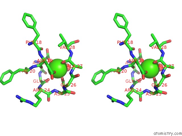
Stereo pair view
Mono view
Stereo pair view
A full contact list of Calcium with other atoms in the Ca binding
site number 1 of Scallop Myosin S1-Adp-P-Pdm in the Actin-Detached Conformation within 5.0Å range:
|
Reference:
D.M.Himmel,
S.Gourinath,
L.Reshetnikova,
Y.Shen,
A.G.Szent-Gyorgyi,
C.Cohen.
Crystallographic Findings on the Internally Uncoupled and Near-Rigor States of Myosin: Further Insights Into the Mechanics of the Motor. Proc.Natl.Acad.Sci.Usa V. 99 12645 2002.
ISSN: ISSN 0027-8424
PubMed: 12297624
DOI: 10.1073/PNAS.202476799
Page generated: Thu Jul 11 11:45:25 2024
ISSN: ISSN 0027-8424
PubMed: 12297624
DOI: 10.1073/PNAS.202476799
Last articles
Zn in 9MJ5Zn in 9HNW
Zn in 9G0L
Zn in 9FNE
Zn in 9DZN
Zn in 9E0I
Zn in 9D32
Zn in 9DAK
Zn in 8ZXC
Zn in 8ZUF