Calcium »
PDB 1l3w-1lkv »
1lhn »
Calcium in PDB 1lhn: Crystal Structure of the N-Terminal Lg-Domain of Shbg in Complex with 5ALPHA-Androstane-3BETA,17ALPHA-Diol
Protein crystallography data
The structure of Crystal Structure of the N-Terminal Lg-Domain of Shbg in Complex with 5ALPHA-Androstane-3BETA,17ALPHA-Diol, PDB code: 1lhn
was solved by
I.Grishkovskaya,
G.V.Avvakumov,
G.L.Hammond,
M.G.Catalano,
Y.A.Muller,
with X-Ray Crystallography technique. A brief refinement statistics is given in the table below:
| Resolution Low / High (Å) | 19.09 / 2.00 |
| Space group | H 3 2 |
| Cell size a, b, c (Å), α, β, γ (°) | 103.900, 103.900, 84.480, 90.00, 90.00, 120.00 |
| R / Rfree (%) | 19.8 / 23.9 |
Other elements in 1lhn:
The structure of Crystal Structure of the N-Terminal Lg-Domain of Shbg in Complex with 5ALPHA-Androstane-3BETA,17ALPHA-Diol also contains other interesting chemical elements:
| Zinc | (Zn) | 1 atom |
Calcium Binding Sites:
The binding sites of Calcium atom in the Crystal Structure of the N-Terminal Lg-Domain of Shbg in Complex with 5ALPHA-Androstane-3BETA,17ALPHA-Diol
(pdb code 1lhn). This binding sites where shown within
5.0 Angstroms radius around Calcium atom.
In total only one binding site of Calcium was determined in the Crystal Structure of the N-Terminal Lg-Domain of Shbg in Complex with 5ALPHA-Androstane-3BETA,17ALPHA-Diol, PDB code: 1lhn:
In total only one binding site of Calcium was determined in the Crystal Structure of the N-Terminal Lg-Domain of Shbg in Complex with 5ALPHA-Androstane-3BETA,17ALPHA-Diol, PDB code: 1lhn:
Calcium binding site 1 out of 1 in 1lhn
Go back to
Calcium binding site 1 out
of 1 in the Crystal Structure of the N-Terminal Lg-Domain of Shbg in Complex with 5ALPHA-Androstane-3BETA,17ALPHA-Diol
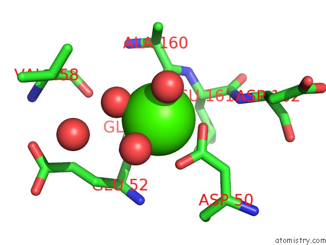
Mono view
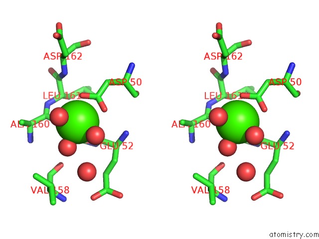
Stereo pair view
Mono view
Stereo pair view
A full contact list of Calcium with other atoms in the Ca binding
site number 1 of Crystal Structure of the N-Terminal Lg-Domain of Shbg in Complex with 5ALPHA-Androstane-3BETA,17ALPHA-Diol within 5.0Å range:
|
Reference:
I.Grishkovskaya,
G.V.Avvakumov,
G.L.Hammond,
M.G.Catalano,
Y.A.Muller.
Steroid Ligands Bind Human Sex Hormone-Binding Globulin in Specific Orientations and Produce Distinct Changes in Protein Conformation J.Biol.Chem. V. 277 32086 2002.
ISSN: ISSN 0021-9258
PubMed: 12065592
DOI: 10.1074/JBC.M203999200
Page generated: Thu Jul 11 11:54:27 2024
ISSN: ISSN 0021-9258
PubMed: 12065592
DOI: 10.1074/JBC.M203999200
Last articles
Zn in 9MJ5Zn in 9HNW
Zn in 9G0L
Zn in 9FNE
Zn in 9DZN
Zn in 9E0I
Zn in 9D32
Zn in 9DAK
Zn in 8ZXC
Zn in 8ZUF