Calcium »
PDB 1l3w-1lkv »
1lkv »
Calcium in PDB 1lkv: Crystal Structure of the Middle and C-Terminal Domains of the Flagellar Rotor Protein Flig
Protein crystallography data
The structure of Crystal Structure of the Middle and C-Terminal Domains of the Flagellar Rotor Protein Flig, PDB code: 1lkv
was solved by
P.N.Brown,
C.P.Hill,
D.F.Blair,
with X-Ray Crystallography technique. A brief refinement statistics is given in the table below:
| Resolution Low / High (Å) | 57.00 / 2.80 |
| Space group | P 64 2 2 |
| Cell size a, b, c (Å), α, β, γ (°) | 113.943, 113.943, 128.826, 90.00, 90.00, 120.00 |
| R / Rfree (%) | 25.5 / 28 |
Calcium Binding Sites:
The binding sites of Calcium atom in the Crystal Structure of the Middle and C-Terminal Domains of the Flagellar Rotor Protein Flig
(pdb code 1lkv). This binding sites where shown within
5.0 Angstroms radius around Calcium atom.
In total 2 binding sites of Calcium where determined in the Crystal Structure of the Middle and C-Terminal Domains of the Flagellar Rotor Protein Flig, PDB code: 1lkv:
Jump to Calcium binding site number: 1; 2;
In total 2 binding sites of Calcium where determined in the Crystal Structure of the Middle and C-Terminal Domains of the Flagellar Rotor Protein Flig, PDB code: 1lkv:
Jump to Calcium binding site number: 1; 2;
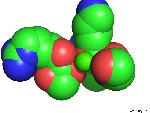
Calcium binding site 1 out of 2 in 1lkv
Go back to
Calcium binding site 1 out
of 2 in the Crystal Structure of the Middle and C-Terminal Domains of the Flagellar Rotor Protein Flig
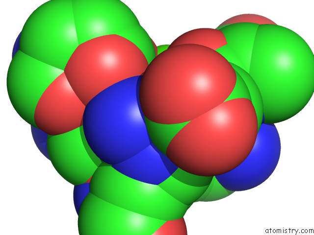
Mono view
Stereo pair view
Mono view
Stereo pair view
A full contact list of Calcium with other atoms in the Ca binding
site number 1 of Crystal Structure of the Middle and C-Terminal Domains of the Flagellar Rotor Protein Flig within 5.0Å range:
|
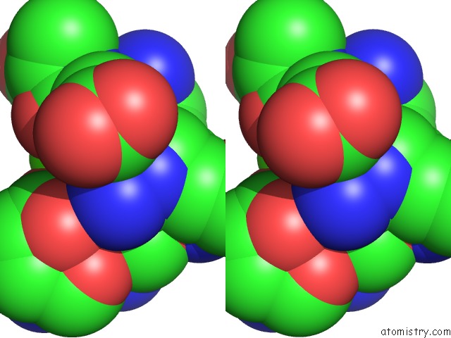
Calcium binding site 2 out of 2 in 1lkv
Go back to
Calcium binding site 2 out
of 2 in the Crystal Structure of the Middle and C-Terminal Domains of the Flagellar Rotor Protein Flig
Mono view

Stereo pair view
Mono view
Stereo pair view
A full contact list of Calcium with other atoms in the Ca binding
site number 2 of Crystal Structure of the Middle and C-Terminal Domains of the Flagellar Rotor Protein Flig within 5.0Å range:
|
Reference:
P.N.Brown,
C.P.Hill,
D.F.Blair.
Crystal Structure of the Middle and C-Terminal Domains of the Flagellar Rotor Protein Flig. Embo J. V. 21 3225 2002.
ISSN: ISSN 0261-4189
PubMed: 12093724
DOI: 10.1093/EMBOJ/CDF332
Page generated: Thu Jul 11 11:55:41 2024
ISSN: ISSN 0261-4189
PubMed: 12093724
DOI: 10.1093/EMBOJ/CDF332
Last articles
Zn in 9MJ5Zn in 9HNW
Zn in 9G0L
Zn in 9FNE
Zn in 9DZN
Zn in 9E0I
Zn in 9D32
Zn in 9DAK
Zn in 8ZXC
Zn in 8ZUF