Calcium »
PDB 1llp-1ltj »
1low »
Calcium in PDB 1low: X-Ray Structure of the H40A Mutant of Ribonuclease T1 Complexed with 3'-Guanosine Monophosphate
Enzymatic activity of X-Ray Structure of the H40A Mutant of Ribonuclease T1 Complexed with 3'-Guanosine Monophosphate
All present enzymatic activity of X-Ray Structure of the H40A Mutant of Ribonuclease T1 Complexed with 3'-Guanosine Monophosphate:
3.1.27.3;
3.1.27.3;
Protein crystallography data
The structure of X-Ray Structure of the H40A Mutant of Ribonuclease T1 Complexed with 3'-Guanosine Monophosphate, PDB code: 1low
was solved by
P.Mignon,
J.Steyaert,
R.Loris,
P.Geerlings,
S.Loverix,
with X-Ray Crystallography technique. A brief refinement statistics is given in the table below:
| Resolution Low / High (Å) | 22.80 / 1.90 |
| Space group | P 21 21 21 |
| Cell size a, b, c (Å), α, β, γ (°) | 40.217, 45.685, 50.136, 90.00, 90.00, 90.00 |
| R / Rfree (%) | 20.6 / 22.9 |
Calcium Binding Sites:
The binding sites of Calcium atom in the X-Ray Structure of the H40A Mutant of Ribonuclease T1 Complexed with 3'-Guanosine Monophosphate
(pdb code 1low). This binding sites where shown within
5.0 Angstroms radius around Calcium atom.
In total only one binding site of Calcium was determined in the X-Ray Structure of the H40A Mutant of Ribonuclease T1 Complexed with 3'-Guanosine Monophosphate, PDB code: 1low:
In total only one binding site of Calcium was determined in the X-Ray Structure of the H40A Mutant of Ribonuclease T1 Complexed with 3'-Guanosine Monophosphate, PDB code: 1low:
Calcium binding site 1 out of 1 in 1low
Go back to
Calcium binding site 1 out
of 1 in the X-Ray Structure of the H40A Mutant of Ribonuclease T1 Complexed with 3'-Guanosine Monophosphate
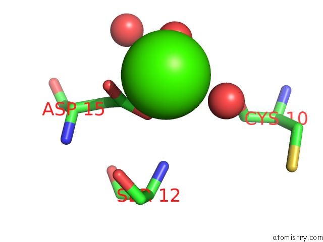
Mono view
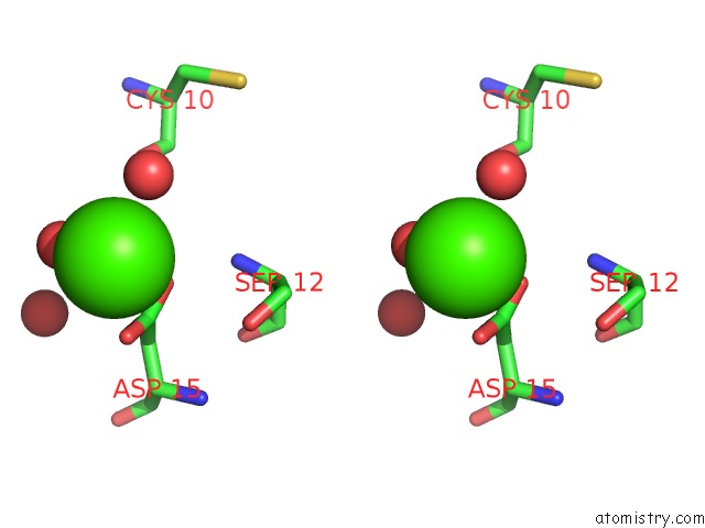
Stereo pair view
Mono view
Stereo pair view
A full contact list of Calcium with other atoms in the Ca binding
site number 1 of X-Ray Structure of the H40A Mutant of Ribonuclease T1 Complexed with 3'-Guanosine Monophosphate within 5.0Å range:
|
Reference:
P.Mignon,
J.Steyaert,
R.Loris,
P.Geerlings,
S.Loverix.
A Nucleophile Activation Dyad in Ribonucleases. A Combined X-Ray Crystallographic/Ab Initio Quantum Chemical Study J.Biol.Chem. V. 277 36770 2002.
ISSN: ISSN 0021-9258
PubMed: 12122018
DOI: 10.1074/JBC.M206461200
Page generated: Thu Jul 11 12:01:40 2024
ISSN: ISSN 0021-9258
PubMed: 12122018
DOI: 10.1074/JBC.M206461200
Last articles
Zn in 9J0NZn in 9J0O
Zn in 9J0P
Zn in 9FJX
Zn in 9EKB
Zn in 9C0F
Zn in 9CAH
Zn in 9CH0
Zn in 9CH3
Zn in 9CH1