Calcium »
PDB 1llp-1ltj »
1lqd »
Calcium in PDB 1lqd: Crystal Structure of Fxa in Complex with 45.
Enzymatic activity of Crystal Structure of Fxa in Complex with 45.
All present enzymatic activity of Crystal Structure of Fxa in Complex with 45.:
3.4.21.6;
3.4.21.6;
Protein crystallography data
The structure of Crystal Structure of Fxa in Complex with 45., PDB code: 1lqd
was solved by
H.A.Schreuder,
P.Loenze,
V.Brachvogel,
A.Liesum,
with X-Ray Crystallography technique. A brief refinement statistics is given in the table below:
| Resolution Low / High (Å) | 8.00 / 2.70 |
| Space group | P 21 21 21 |
| Cell size a, b, c (Å), α, β, γ (°) | 56.659, 72.398, 78.486, 90.00, 90.00, 90.00 |
| R / Rfree (%) | 16.1 / 29.5 |
Calcium Binding Sites:
The binding sites of Calcium atom in the Crystal Structure of Fxa in Complex with 45.
(pdb code 1lqd). This binding sites where shown within
5.0 Angstroms radius around Calcium atom.
In total only one binding site of Calcium was determined in the Crystal Structure of Fxa in Complex with 45., PDB code: 1lqd:
In total only one binding site of Calcium was determined in the Crystal Structure of Fxa in Complex with 45., PDB code: 1lqd:
Calcium binding site 1 out of 1 in 1lqd
Go back to
Calcium binding site 1 out
of 1 in the Crystal Structure of Fxa in Complex with 45.
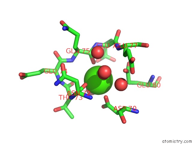
Mono view
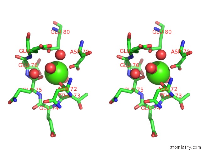
Stereo pair view
Mono view
Stereo pair view
A full contact list of Calcium with other atoms in the Ca binding
site number 1 of Crystal Structure of Fxa in Complex with 45. within 5.0Å range:
|
Reference:
H.Matter,
E.Defossa,
U.Heinelt,
P.M.Blohm,
D.Schneider,
A.Mueller,
S.Herok,
H.A.Schreuder,
A.Liesum,
V.Brachvogel,
P.Loenze,
A.Walser,
F.Al-Obeidi,
P.Wildgoose.
Design and Quantitative Structure-Activity Relationship of 3-Amidinobenzyl-1H-Indole-2-Carboxamides As Potent, Nonchiral, and Selective Inhibitors of Blood Coagulation Factor Xa J.Med.Chem. V. 45 2749 2002.
ISSN: ISSN 0022-2623
PubMed: 12061878
DOI: 10.1021/JM0111346
Page generated: Thu Jul 11 12:03:07 2024
ISSN: ISSN 0022-2623
PubMed: 12061878
DOI: 10.1021/JM0111346
Last articles
Zn in 9J0NZn in 9J0O
Zn in 9J0P
Zn in 9FJX
Zn in 9EKB
Zn in 9C0F
Zn in 9CAH
Zn in 9CH0
Zn in 9CH3
Zn in 9CH1