Calcium »
PDB 1llp-1ltj »
1lqv »
Calcium in PDB 1lqv: Crystal Structure of the Endothelial Protein C Receptor with Phospholipid in the Groove in Complex with Gla Domain of Protein C.
Protein crystallography data
The structure of Crystal Structure of the Endothelial Protein C Receptor with Phospholipid in the Groove in Complex with Gla Domain of Protein C., PDB code: 1lqv
was solved by
V.Oganesyan,
N.Oganesyan,
S.Terzyan,
Q.Dongfeng,
Z.Dauter,
N.L.Esmon,
C.T.Esmon,
with X-Ray Crystallography technique. A brief refinement statistics is given in the table below:
| Resolution Low / High (Å) | 12.00 / 1.60 |
| Space group | P 1 21 1 |
| Cell size a, b, c (Å), α, β, γ (°) | 59.220, 62.360, 71.030, 90.00, 101.81, 90.00 |
| R / Rfree (%) | 18.9 / 22.2 |
Calcium Binding Sites:
Pages:
>>> Page 1 <<< Page 2, Binding sites: 11 - 14;Binding sites:
The binding sites of Calcium atom in the Crystal Structure of the Endothelial Protein C Receptor with Phospholipid in the Groove in Complex with Gla Domain of Protein C. (pdb code 1lqv). This binding sites where shown within 5.0 Angstroms radius around Calcium atom.In total 14 binding sites of Calcium where determined in the Crystal Structure of the Endothelial Protein C Receptor with Phospholipid in the Groove in Complex with Gla Domain of Protein C., PDB code: 1lqv:
Jump to Calcium binding site number: 1; 2; 3; 4; 5; 6; 7; 8; 9; 10;
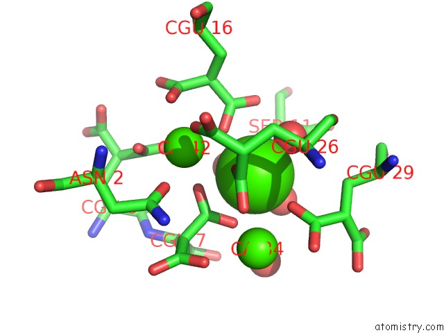
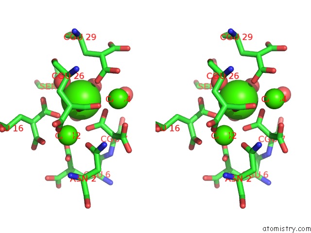
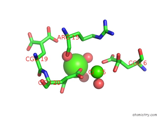
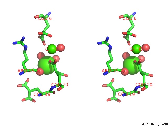
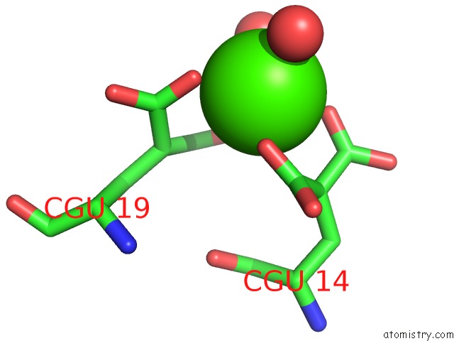
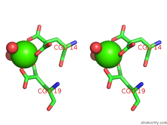
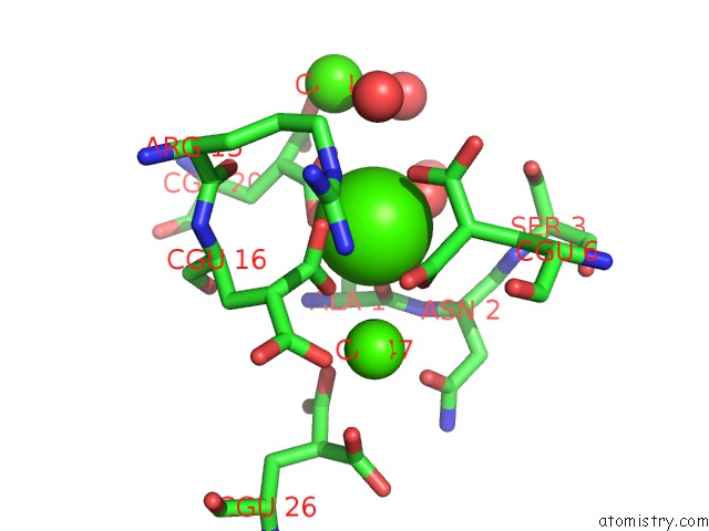
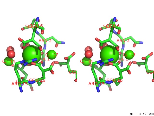
Calcium binding site 1 out of 14 in 1lqv
Go back to
Calcium binding site 1 out
of 14 in the Crystal Structure of the Endothelial Protein C Receptor with Phospholipid in the Groove in Complex with Gla Domain of Protein C.
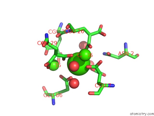
Mono view
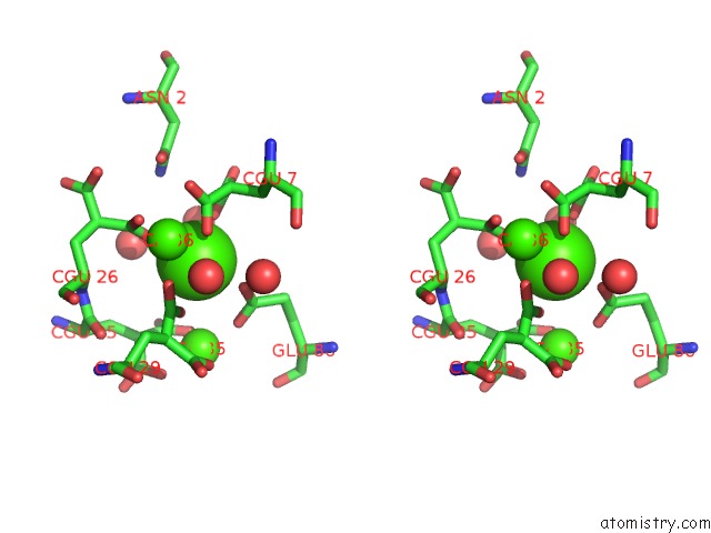
Stereo pair view
Mono view
Stereo pair view
A full contact list of Calcium with other atoms in the Ca binding
site number 1 of Crystal Structure of the Endothelial Protein C Receptor with Phospholipid in the Groove in Complex with Gla Domain of Protein C. within 5.0Å range:
|
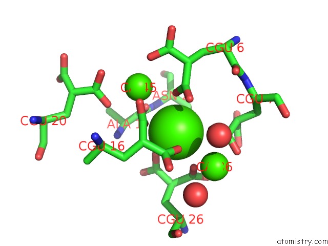
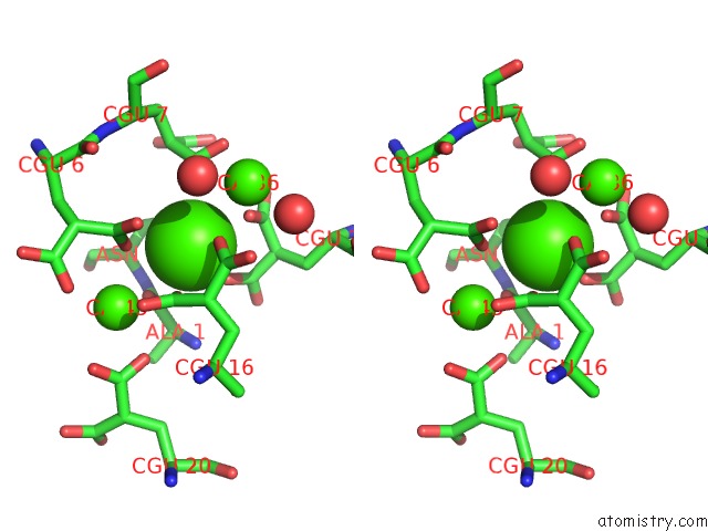
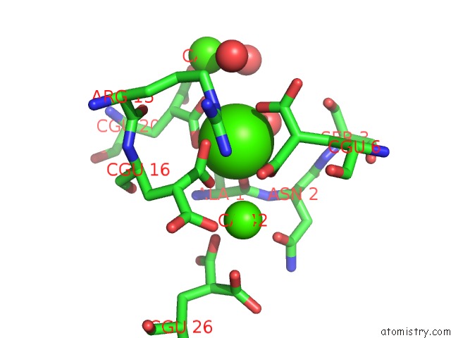
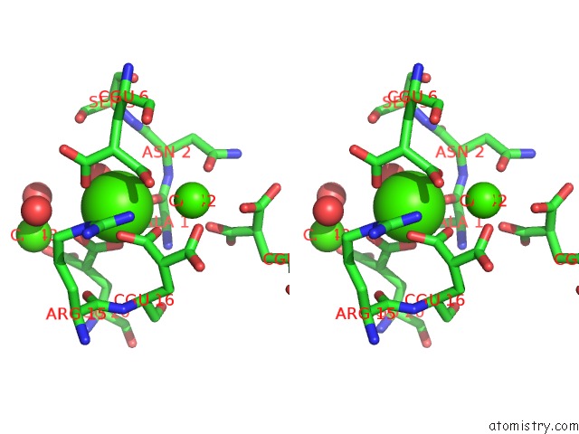
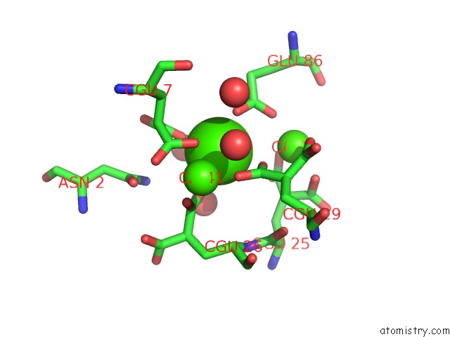
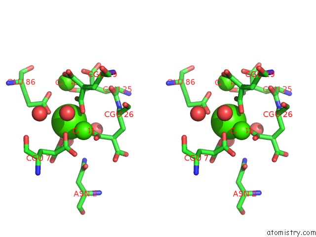
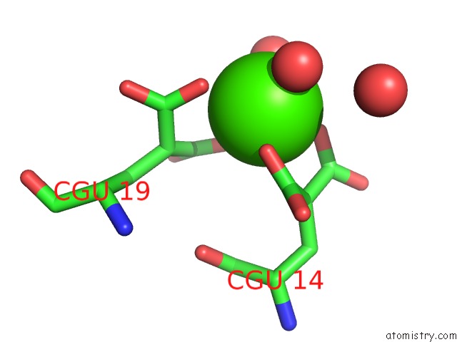
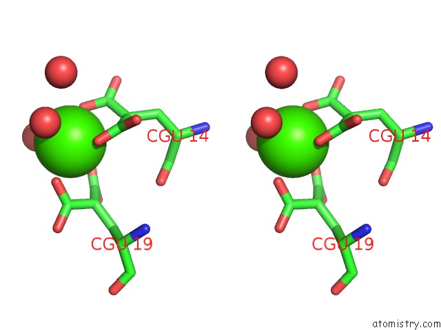
Calcium binding site 2 out of 14 in 1lqv
Go back to
Calcium binding site 2 out
of 14 in the Crystal Structure of the Endothelial Protein C Receptor with Phospholipid in the Groove in Complex with Gla Domain of Protein C.
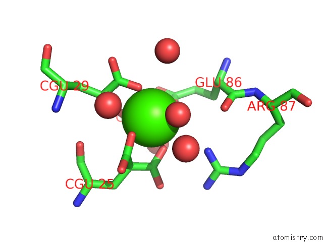
Mono view
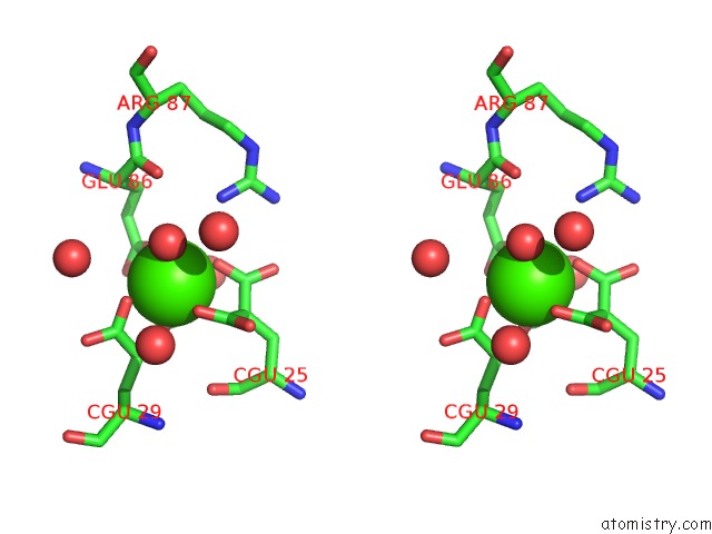
Stereo pair view
Mono view
Stereo pair view
A full contact list of Calcium with other atoms in the Ca binding
site number 2 of Crystal Structure of the Endothelial Protein C Receptor with Phospholipid in the Groove in Complex with Gla Domain of Protein C. within 5.0Å range:
|
Calcium binding site 3 out of 14 in 1lqv
Go back to
Calcium binding site 3 out
of 14 in the Crystal Structure of the Endothelial Protein C Receptor with Phospholipid in the Groove in Complex with Gla Domain of Protein C.
Mono view
Stereo pair view
Mono view
Stereo pair view
A full contact list of Calcium with other atoms in the Ca binding
site number 3 of Crystal Structure of the Endothelial Protein C Receptor with Phospholipid in the Groove in Complex with Gla Domain of Protein C. within 5.0Å range:
|
Calcium binding site 4 out of 14 in 1lqv
Go back to
Calcium binding site 4 out
of 14 in the Crystal Structure of the Endothelial Protein C Receptor with Phospholipid in the Groove in Complex with Gla Domain of Protein C.
Mono view
Stereo pair view
Mono view
Stereo pair view
A full contact list of Calcium with other atoms in the Ca binding
site number 4 of Crystal Structure of the Endothelial Protein C Receptor with Phospholipid in the Groove in Complex with Gla Domain of Protein C. within 5.0Å range:
|
Calcium binding site 5 out of 14 in 1lqv
Go back to
Calcium binding site 5 out
of 14 in the Crystal Structure of the Endothelial Protein C Receptor with Phospholipid in the Groove in Complex with Gla Domain of Protein C.
Mono view
Stereo pair view
Mono view
Stereo pair view
A full contact list of Calcium with other atoms in the Ca binding
site number 5 of Crystal Structure of the Endothelial Protein C Receptor with Phospholipid in the Groove in Complex with Gla Domain of Protein C. within 5.0Å range:
|
Calcium binding site 6 out of 14 in 1lqv
Go back to
Calcium binding site 6 out
of 14 in the Crystal Structure of the Endothelial Protein C Receptor with Phospholipid in the Groove in Complex with Gla Domain of Protein C.
Mono view
Stereo pair view
Mono view
Stereo pair view
A full contact list of Calcium with other atoms in the Ca binding
site number 6 of Crystal Structure of the Endothelial Protein C Receptor with Phospholipid in the Groove in Complex with Gla Domain of Protein C. within 5.0Å range:
|
Calcium binding site 7 out of 14 in 1lqv
Go back to
Calcium binding site 7 out
of 14 in the Crystal Structure of the Endothelial Protein C Receptor with Phospholipid in the Groove in Complex with Gla Domain of Protein C.
Mono view
Stereo pair view
Mono view
Stereo pair view
A full contact list of Calcium with other atoms in the Ca binding
site number 7 of Crystal Structure of the Endothelial Protein C Receptor with Phospholipid in the Groove in Complex with Gla Domain of Protein C. within 5.0Å range:
|
Calcium binding site 8 out of 14 in 1lqv
Go back to
Calcium binding site 8 out
of 14 in the Crystal Structure of the Endothelial Protein C Receptor with Phospholipid in the Groove in Complex with Gla Domain of Protein C.
Mono view
Stereo pair view
Mono view
Stereo pair view
A full contact list of Calcium with other atoms in the Ca binding
site number 8 of Crystal Structure of the Endothelial Protein C Receptor with Phospholipid in the Groove in Complex with Gla Domain of Protein C. within 5.0Å range:
|
Calcium binding site 9 out of 14 in 1lqv
Go back to
Calcium binding site 9 out
of 14 in the Crystal Structure of the Endothelial Protein C Receptor with Phospholipid in the Groove in Complex with Gla Domain of Protein C.
Mono view
Stereo pair view
Mono view
Stereo pair view
A full contact list of Calcium with other atoms in the Ca binding
site number 9 of Crystal Structure of the Endothelial Protein C Receptor with Phospholipid in the Groove in Complex with Gla Domain of Protein C. within 5.0Å range:
|
Calcium binding site 10 out of 14 in 1lqv
Go back to
Calcium binding site 10 out
of 14 in the Crystal Structure of the Endothelial Protein C Receptor with Phospholipid in the Groove in Complex with Gla Domain of Protein C.
Mono view
Stereo pair view
Mono view
Stereo pair view
A full contact list of Calcium with other atoms in the Ca binding
site number 10 of Crystal Structure of the Endothelial Protein C Receptor with Phospholipid in the Groove in Complex with Gla Domain of Protein C. within 5.0Å range:
|
Reference:
V.Oganesyan,
N.Oganesyan,
S.Terzyan,
D.Qu,
Z.Dauter,
N.L.Esmon,
C.T.Esmon.
The Crystal Structure of the Endothelial Protein C Receptor and A Bound Phospholipid. J.Biol.Chem. V. 277 24851 2002.
ISSN: ISSN 0021-9258
PubMed: 12034704
DOI: 10.1074/JBC.C200163200
Page generated: Thu Jul 11 12:03:19 2024
ISSN: ISSN 0021-9258
PubMed: 12034704
DOI: 10.1074/JBC.C200163200
Last articles
Zn in 9J0NZn in 9J0O
Zn in 9J0P
Zn in 9FJX
Zn in 9EKB
Zn in 9C0F
Zn in 9CAH
Zn in 9CH0
Zn in 9CH3
Zn in 9CH1