Calcium »
PDB 1lu1-1m8v »
1lv8 »
Calcium in PDB 1lv8: Crystal Structure of Calf Spleen Purine Nucleoside Phosphorylase in A New Space Group with Full Trimer in the Asymmetric Unit
Enzymatic activity of Crystal Structure of Calf Spleen Purine Nucleoside Phosphorylase in A New Space Group with Full Trimer in the Asymmetric Unit
All present enzymatic activity of Crystal Structure of Calf Spleen Purine Nucleoside Phosphorylase in A New Space Group with Full Trimer in the Asymmetric Unit:
2.4.2.1;
2.4.2.1;
Protein crystallography data
The structure of Crystal Structure of Calf Spleen Purine Nucleoside Phosphorylase in A New Space Group with Full Trimer in the Asymmetric Unit, PDB code: 1lv8
was solved by
A.Bzowska,
G.Koellner,
B.Wielgus-Kutrowska,
A.Stroh,
G.Raszewski,
A.Holy,
T.Steiner,
J.Frank,
with X-Ray Crystallography technique. A brief refinement statistics is given in the table below:
| Resolution Low / High (Å) | 19.85 / 2.30 |
| Space group | P 21 21 21 |
| Cell size a, b, c (Å), α, β, γ (°) | 79.140, 134.235, 177.148, 90.00, 90.00, 90.00 |
| R / Rfree (%) | 18.9 / 25.8 |
Calcium Binding Sites:
The binding sites of Calcium atom in the Crystal Structure of Calf Spleen Purine Nucleoside Phosphorylase in A New Space Group with Full Trimer in the Asymmetric Unit
(pdb code 1lv8). This binding sites where shown within
5.0 Angstroms radius around Calcium atom.
In total 6 binding sites of Calcium where determined in the Crystal Structure of Calf Spleen Purine Nucleoside Phosphorylase in A New Space Group with Full Trimer in the Asymmetric Unit, PDB code: 1lv8:
Jump to Calcium binding site number: 1; 2; 3; 4; 5; 6;
In total 6 binding sites of Calcium where determined in the Crystal Structure of Calf Spleen Purine Nucleoside Phosphorylase in A New Space Group with Full Trimer in the Asymmetric Unit, PDB code: 1lv8:
Jump to Calcium binding site number: 1; 2; 3; 4; 5; 6;
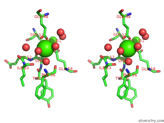
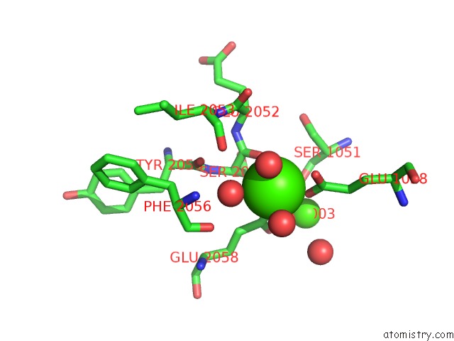
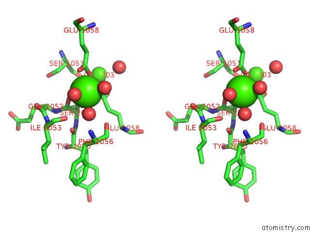
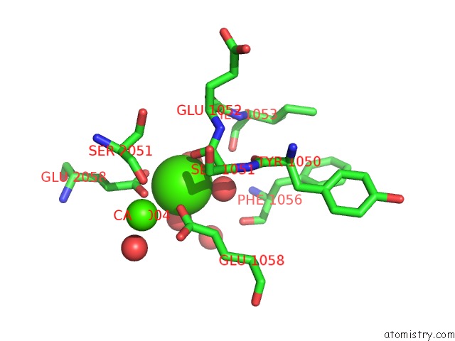
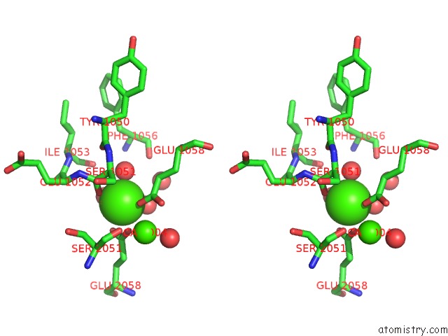
Calcium binding site 1 out of 6 in 1lv8
Go back to
Calcium binding site 1 out
of 6 in the Crystal Structure of Calf Spleen Purine Nucleoside Phosphorylase in A New Space Group with Full Trimer in the Asymmetric Unit
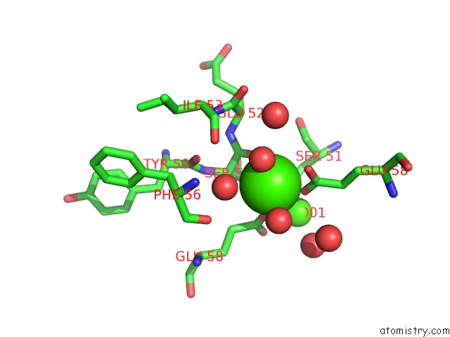
Mono view
Stereo pair view
Mono view
Stereo pair view
A full contact list of Calcium with other atoms in the Ca binding
site number 1 of Crystal Structure of Calf Spleen Purine Nucleoside Phosphorylase in A New Space Group with Full Trimer in the Asymmetric Unit within 5.0Å range:
|
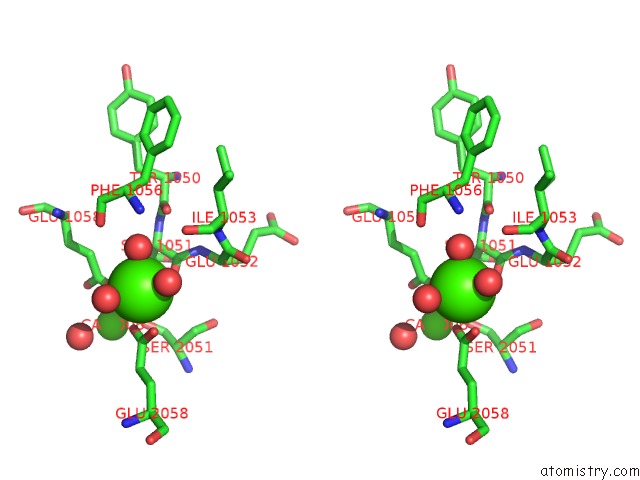
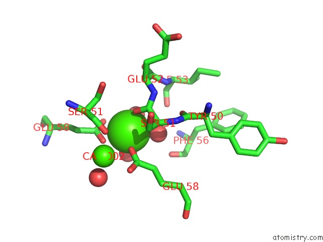
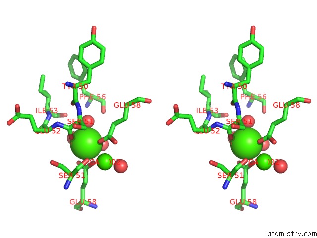
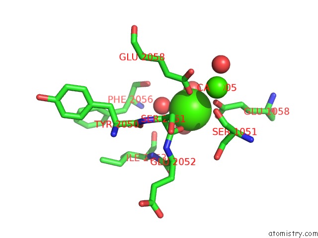
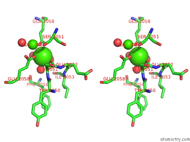
Calcium binding site 2 out of 6 in 1lv8
Go back to
Calcium binding site 2 out
of 6 in the Crystal Structure of Calf Spleen Purine Nucleoside Phosphorylase in A New Space Group with Full Trimer in the Asymmetric Unit
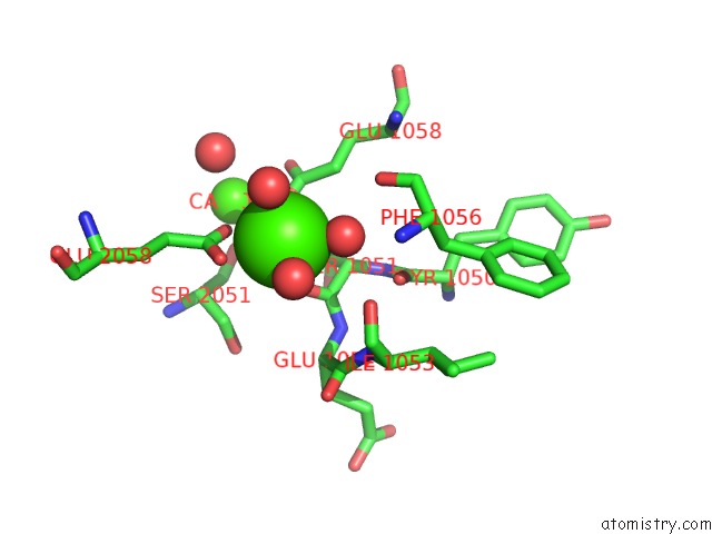
Mono view
Stereo pair view
Mono view
Stereo pair view
A full contact list of Calcium with other atoms in the Ca binding
site number 2 of Crystal Structure of Calf Spleen Purine Nucleoside Phosphorylase in A New Space Group with Full Trimer in the Asymmetric Unit within 5.0Å range:
|
Calcium binding site 3 out of 6 in 1lv8
Go back to
Calcium binding site 3 out
of 6 in the Crystal Structure of Calf Spleen Purine Nucleoside Phosphorylase in A New Space Group with Full Trimer in the Asymmetric Unit
Mono view
Stereo pair view
Mono view
Stereo pair view
A full contact list of Calcium with other atoms in the Ca binding
site number 3 of Crystal Structure of Calf Spleen Purine Nucleoside Phosphorylase in A New Space Group with Full Trimer in the Asymmetric Unit within 5.0Å range:
|
Calcium binding site 4 out of 6 in 1lv8
Go back to
Calcium binding site 4 out
of 6 in the Crystal Structure of Calf Spleen Purine Nucleoside Phosphorylase in A New Space Group with Full Trimer in the Asymmetric Unit
Mono view
Stereo pair view
Mono view
Stereo pair view
A full contact list of Calcium with other atoms in the Ca binding
site number 4 of Crystal Structure of Calf Spleen Purine Nucleoside Phosphorylase in A New Space Group with Full Trimer in the Asymmetric Unit within 5.0Å range:
|
Calcium binding site 5 out of 6 in 1lv8
Go back to
Calcium binding site 5 out
of 6 in the Crystal Structure of Calf Spleen Purine Nucleoside Phosphorylase in A New Space Group with Full Trimer in the Asymmetric Unit
Mono view
Stereo pair view
Mono view
Stereo pair view
A full contact list of Calcium with other atoms in the Ca binding
site number 5 of Crystal Structure of Calf Spleen Purine Nucleoside Phosphorylase in A New Space Group with Full Trimer in the Asymmetric Unit within 5.0Å range:
|
Calcium binding site 6 out of 6 in 1lv8
Go back to
Calcium binding site 6 out
of 6 in the Crystal Structure of Calf Spleen Purine Nucleoside Phosphorylase in A New Space Group with Full Trimer in the Asymmetric Unit
Mono view
Stereo pair view
Mono view
Stereo pair view
A full contact list of Calcium with other atoms in the Ca binding
site number 6 of Crystal Structure of Calf Spleen Purine Nucleoside Phosphorylase in A New Space Group with Full Trimer in the Asymmetric Unit within 5.0Å range:
|
Reference:
A.Bzowska,
G.Koellner,
B.Wielgus-Kutrowska,
A.Stroh,
G.Raszewski,
A.Holy,
T.Steiner,
J.Frank.
Crystal Structure of Calf Spleen Purine Nucleoside Phosphorylase with Two Full Trimers in the Asymmetric Unit: Important Implications For the Mechanism of Catalysis J.Mol.Biol. V. 342 1015 2004.
ISSN: ISSN 0022-2836
PubMed: 15342253
DOI: 10.1016/J.JMB.2004.07.017
Page generated: Thu Jul 11 12:07:08 2024
ISSN: ISSN 0022-2836
PubMed: 15342253
DOI: 10.1016/J.JMB.2004.07.017
Last articles
Zn in 9JYWZn in 9IR4
Zn in 9IR3
Zn in 9GMX
Zn in 9GMW
Zn in 9JEJ
Zn in 9ERF
Zn in 9ERE
Zn in 9EGV
Zn in 9EGW