Calcium »
PDB 1lu1-1m8v »
1lvc »
Calcium in PDB 1lvc: Crystal Structure of the Adenylyl Cyclase Domain of Anthrax Edema Factor (Ef) in Complex with Calmodulin and 2' Deoxy, 3' Anthraniloyl Atp
Enzymatic activity of Crystal Structure of the Adenylyl Cyclase Domain of Anthrax Edema Factor (Ef) in Complex with Calmodulin and 2' Deoxy, 3' Anthraniloyl Atp
All present enzymatic activity of Crystal Structure of the Adenylyl Cyclase Domain of Anthrax Edema Factor (Ef) in Complex with Calmodulin and 2' Deoxy, 3' Anthraniloyl Atp:
4.6.1.1;
4.6.1.1;
Protein crystallography data
The structure of Crystal Structure of the Adenylyl Cyclase Domain of Anthrax Edema Factor (Ef) in Complex with Calmodulin and 2' Deoxy, 3' Anthraniloyl Atp, PDB code: 1lvc
was solved by
Y.Shen,
Y.-S.Lee,
S.Soelaiman,
P.Bergson,
D.Lu,
A.Chen,
K.Beckingham,
Z.Grabarek,
M.Mrksich,
W.-J.Tang,
with X-Ray Crystallography technique. A brief refinement statistics is given in the table below:
| Resolution Low / High (Å) | 29.96 / 3.60 |
| Space group | I 2 2 2 |
| Cell size a, b, c (Å), α, β, γ (°) | 116.924, 167.918, 341.743, 90.00, 90.00, 90.00 |
| R / Rfree (%) | 28.1 / 30.7 |
Other elements in 1lvc:
The structure of Crystal Structure of the Adenylyl Cyclase Domain of Anthrax Edema Factor (Ef) in Complex with Calmodulin and 2' Deoxy, 3' Anthraniloyl Atp also contains other interesting chemical elements:
| Ytterbium | (Yb) | 3 atoms |
Calcium Binding Sites:
The binding sites of Calcium atom in the Crystal Structure of the Adenylyl Cyclase Domain of Anthrax Edema Factor (Ef) in Complex with Calmodulin and 2' Deoxy, 3' Anthraniloyl Atp
(pdb code 1lvc). This binding sites where shown within
5.0 Angstroms radius around Calcium atom.
In total 6 binding sites of Calcium where determined in the Crystal Structure of the Adenylyl Cyclase Domain of Anthrax Edema Factor (Ef) in Complex with Calmodulin and 2' Deoxy, 3' Anthraniloyl Atp, PDB code: 1lvc:
Jump to Calcium binding site number: 1; 2; 3; 4; 5; 6;
In total 6 binding sites of Calcium where determined in the Crystal Structure of the Adenylyl Cyclase Domain of Anthrax Edema Factor (Ef) in Complex with Calmodulin and 2' Deoxy, 3' Anthraniloyl Atp, PDB code: 1lvc:
Jump to Calcium binding site number: 1; 2; 3; 4; 5; 6;
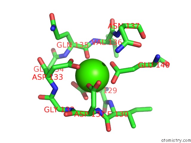
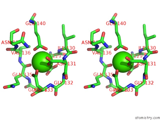
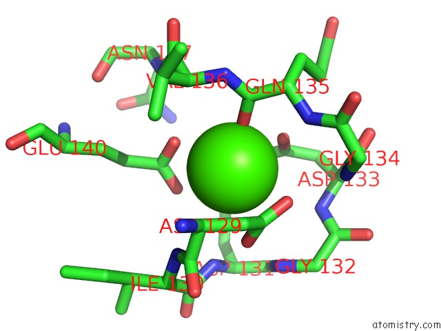
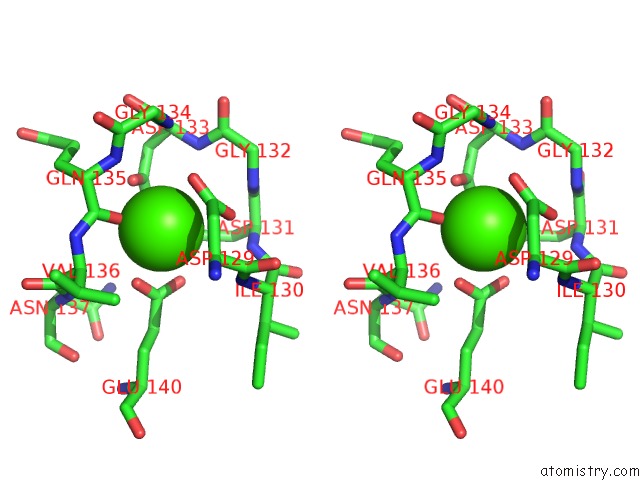
Calcium binding site 1 out of 6 in 1lvc
Go back to
Calcium binding site 1 out
of 6 in the Crystal Structure of the Adenylyl Cyclase Domain of Anthrax Edema Factor (Ef) in Complex with Calmodulin and 2' Deoxy, 3' Anthraniloyl Atp
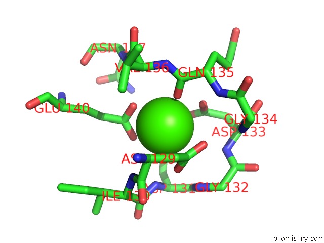
Mono view
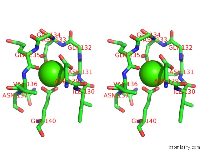
Stereo pair view
Mono view
Stereo pair view
A full contact list of Calcium with other atoms in the Ca binding
site number 1 of Crystal Structure of the Adenylyl Cyclase Domain of Anthrax Edema Factor (Ef) in Complex with Calmodulin and 2' Deoxy, 3' Anthraniloyl Atp within 5.0Å range:
|
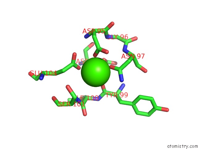
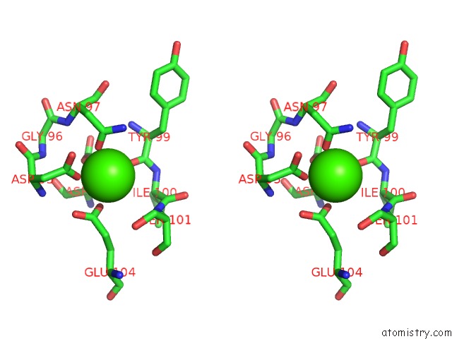
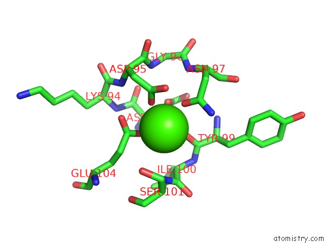
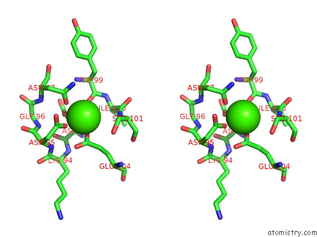
Calcium binding site 2 out of 6 in 1lvc
Go back to
Calcium binding site 2 out
of 6 in the Crystal Structure of the Adenylyl Cyclase Domain of Anthrax Edema Factor (Ef) in Complex with Calmodulin and 2' Deoxy, 3' Anthraniloyl Atp
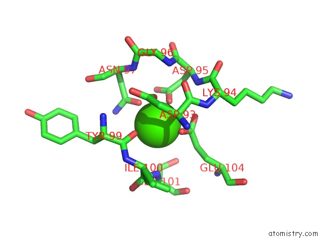
Mono view
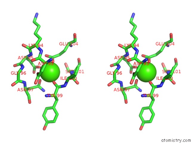
Stereo pair view
Mono view
Stereo pair view
A full contact list of Calcium with other atoms in the Ca binding
site number 2 of Crystal Structure of the Adenylyl Cyclase Domain of Anthrax Edema Factor (Ef) in Complex with Calmodulin and 2' Deoxy, 3' Anthraniloyl Atp within 5.0Å range:
|
Calcium binding site 3 out of 6 in 1lvc
Go back to
Calcium binding site 3 out
of 6 in the Crystal Structure of the Adenylyl Cyclase Domain of Anthrax Edema Factor (Ef) in Complex with Calmodulin and 2' Deoxy, 3' Anthraniloyl Atp
Mono view
Stereo pair view
Mono view
Stereo pair view
A full contact list of Calcium with other atoms in the Ca binding
site number 3 of Crystal Structure of the Adenylyl Cyclase Domain of Anthrax Edema Factor (Ef) in Complex with Calmodulin and 2' Deoxy, 3' Anthraniloyl Atp within 5.0Å range:
|
Calcium binding site 4 out of 6 in 1lvc
Go back to
Calcium binding site 4 out
of 6 in the Crystal Structure of the Adenylyl Cyclase Domain of Anthrax Edema Factor (Ef) in Complex with Calmodulin and 2' Deoxy, 3' Anthraniloyl Atp
Mono view
Stereo pair view
Mono view
Stereo pair view
A full contact list of Calcium with other atoms in the Ca binding
site number 4 of Crystal Structure of the Adenylyl Cyclase Domain of Anthrax Edema Factor (Ef) in Complex with Calmodulin and 2' Deoxy, 3' Anthraniloyl Atp within 5.0Å range:
|
Calcium binding site 5 out of 6 in 1lvc
Go back to
Calcium binding site 5 out
of 6 in the Crystal Structure of the Adenylyl Cyclase Domain of Anthrax Edema Factor (Ef) in Complex with Calmodulin and 2' Deoxy, 3' Anthraniloyl Atp
Mono view
Stereo pair view
Mono view
Stereo pair view
A full contact list of Calcium with other atoms in the Ca binding
site number 5 of Crystal Structure of the Adenylyl Cyclase Domain of Anthrax Edema Factor (Ef) in Complex with Calmodulin and 2' Deoxy, 3' Anthraniloyl Atp within 5.0Å range:
|
Calcium binding site 6 out of 6 in 1lvc
Go back to
Calcium binding site 6 out
of 6 in the Crystal Structure of the Adenylyl Cyclase Domain of Anthrax Edema Factor (Ef) in Complex with Calmodulin and 2' Deoxy, 3' Anthraniloyl Atp
Mono view
Stereo pair view
Mono view
Stereo pair view
A full contact list of Calcium with other atoms in the Ca binding
site number 6 of Crystal Structure of the Adenylyl Cyclase Domain of Anthrax Edema Factor (Ef) in Complex with Calmodulin and 2' Deoxy, 3' Anthraniloyl Atp within 5.0Å range:
|
Reference:
Y.Shen,
Y.-S.Lee,
S.Soelaiman,
P.Bergson,
D.Lu,
A.Chen,
K.Beckingham,
Z.Grabarek,
M.Mrksich,
W.-J.Tang.
Physiological Calcium Concentrations Regulate Calmodulin Binding and Catalysis of Adenylyl Cyclase Exotoxins Embo J. V. 21 6721 2002.
ISSN: ISSN 0261-4189
PubMed: 12485993
DOI: 10.1093/EMBOJ/CDF681
Page generated: Mon Jul 7 17:00:45 2025
ISSN: ISSN 0261-4189
PubMed: 12485993
DOI: 10.1093/EMBOJ/CDF681
Last articles
Ca in 7KGZCa in 7KGQ
Ca in 7KFA
Ca in 7KEV
Ca in 7KBH
Ca in 7KDP
Ca in 7KDD
Ca in 7KBT
Ca in 7KBR
Ca in 7KBJ