Calcium »
PDB 1lu1-1m8v »
1lvn »
Calcium in PDB 1lvn: Crystal Structure of E. Coli Amine Oxidase Complexed with Tranylcypromine
Enzymatic activity of Crystal Structure of E. Coli Amine Oxidase Complexed with Tranylcypromine
All present enzymatic activity of Crystal Structure of E. Coli Amine Oxidase Complexed with Tranylcypromine:
1.4.3.6;
1.4.3.6;
Protein crystallography data
The structure of Crystal Structure of E. Coli Amine Oxidase Complexed with Tranylcypromine, PDB code: 1lvn
was solved by
C.M.Wilmot,
S.E.Phillips,
with X-Ray Crystallography technique. A brief refinement statistics is given in the table below:
| Resolution Low / High (Å) | 20.00 / 2.40 |
| Space group | P 21 21 21 |
| Cell size a, b, c (Å), α, β, γ (°) | 135.236, 166.482, 79.628, 90.00, 90.00, 90.00 |
| R / Rfree (%) | 18.5 / 22.9 |
Other elements in 1lvn:
The structure of Crystal Structure of E. Coli Amine Oxidase Complexed with Tranylcypromine also contains other interesting chemical elements:
| Copper | (Cu) | 2 atoms |
Calcium Binding Sites:
The binding sites of Calcium atom in the Crystal Structure of E. Coli Amine Oxidase Complexed with Tranylcypromine
(pdb code 1lvn). This binding sites where shown within
5.0 Angstroms radius around Calcium atom.
In total 4 binding sites of Calcium where determined in the Crystal Structure of E. Coli Amine Oxidase Complexed with Tranylcypromine, PDB code: 1lvn:
Jump to Calcium binding site number: 1; 2; 3; 4;
In total 4 binding sites of Calcium where determined in the Crystal Structure of E. Coli Amine Oxidase Complexed with Tranylcypromine, PDB code: 1lvn:
Jump to Calcium binding site number: 1; 2; 3; 4;
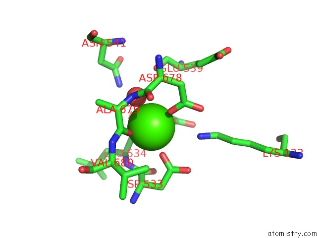
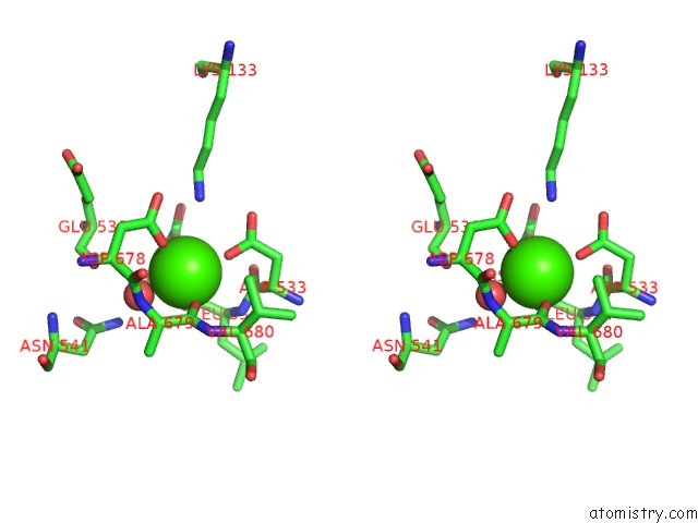
Calcium binding site 1 out of 4 in 1lvn
Go back to
Calcium binding site 1 out
of 4 in the Crystal Structure of E. Coli Amine Oxidase Complexed with Tranylcypromine
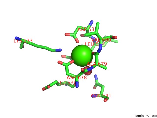
Mono view
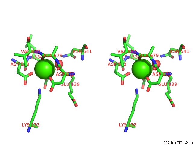
Stereo pair view
Mono view
Stereo pair view
A full contact list of Calcium with other atoms in the Ca binding
site number 1 of Crystal Structure of E. Coli Amine Oxidase Complexed with Tranylcypromine within 5.0Å range:
|
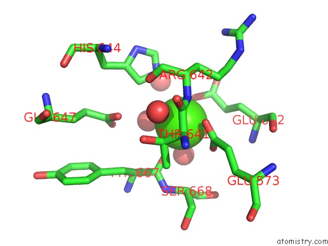
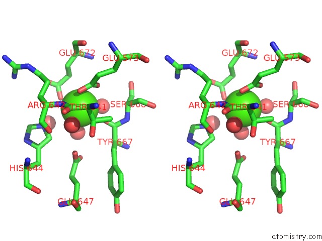
Calcium binding site 2 out of 4 in 1lvn
Go back to
Calcium binding site 2 out
of 4 in the Crystal Structure of E. Coli Amine Oxidase Complexed with Tranylcypromine
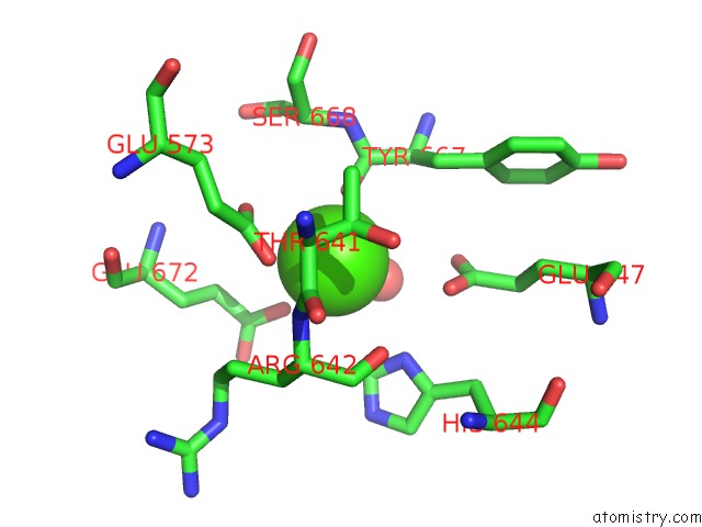
Mono view
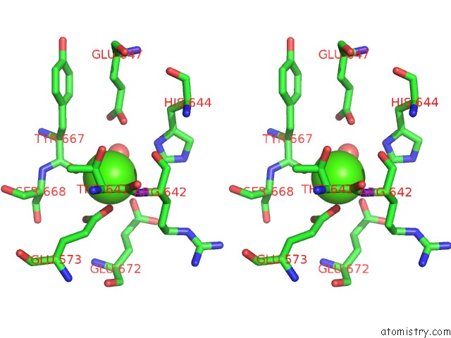
Stereo pair view
Mono view
Stereo pair view
A full contact list of Calcium with other atoms in the Ca binding
site number 2 of Crystal Structure of E. Coli Amine Oxidase Complexed with Tranylcypromine within 5.0Å range:
|
Calcium binding site 3 out of 4 in 1lvn
Go back to
Calcium binding site 3 out
of 4 in the Crystal Structure of E. Coli Amine Oxidase Complexed with Tranylcypromine
Mono view
Stereo pair view
Mono view
Stereo pair view
A full contact list of Calcium with other atoms in the Ca binding
site number 3 of Crystal Structure of E. Coli Amine Oxidase Complexed with Tranylcypromine within 5.0Å range:
|
Calcium binding site 4 out of 4 in 1lvn
Go back to
Calcium binding site 4 out
of 4 in the Crystal Structure of E. Coli Amine Oxidase Complexed with Tranylcypromine
Mono view
Stereo pair view
Mono view
Stereo pair view
A full contact list of Calcium with other atoms in the Ca binding
site number 4 of Crystal Structure of E. Coli Amine Oxidase Complexed with Tranylcypromine within 5.0Å range:
|
Reference:
C.M.Wilmot,
C.G.Saysell,
A.Blessington,
D.A.Conn,
C.R.Kurtis,
M.J.Mcpherson,
P.F.Knowles,
S.E.Phillips.
Medical Implications From the Crystal Structure of A Copper-Containing Amine Oxidase Complexed with the Antidepressant Drug Tranylcypromine. Febs Lett. V. 576 301 2004.
ISSN: ISSN 0014-5793
PubMed: 15498552
DOI: 10.1016/J.FEBSLET.2004.09.031
Page generated: Thu Jul 11 12:07:08 2024
ISSN: ISSN 0014-5793
PubMed: 15498552
DOI: 10.1016/J.FEBSLET.2004.09.031
Last articles
Zn in 9MJ5Zn in 9HNW
Zn in 9G0L
Zn in 9FNE
Zn in 9DZN
Zn in 9E0I
Zn in 9D32
Zn in 9DAK
Zn in 8ZXC
Zn in 8ZUF