Calcium »
PDB 1lu1-1m8v »
1m5x »
Calcium in PDB 1m5x: Crystal Structure of the Homing Endonuclease I-Msoi Bound to Its Dna Substrate
Protein crystallography data
The structure of Crystal Structure of the Homing Endonuclease I-Msoi Bound to Its Dna Substrate, PDB code: 1m5x
was solved by
B.Chevalier,
M.Turmel,
C.Lemieux,
R.J.Monnat,
B.L.Stoddard,
with X-Ray Crystallography technique. A brief refinement statistics is given in the table below:
| Resolution Low / High (Å) | 19.96 / 2.25 |
| Space group | P 1 |
| Cell size a, b, c (Å), α, β, γ (°) | 41.510, 42.210, 71.320, 73.34, 73.17, 71.09 |
| R / Rfree (%) | 21.9 / 25.5 |
Calcium Binding Sites:
The binding sites of Calcium atom in the Crystal Structure of the Homing Endonuclease I-Msoi Bound to Its Dna Substrate
(pdb code 1m5x). This binding sites where shown within
5.0 Angstroms radius around Calcium atom.
In total 3 binding sites of Calcium where determined in the Crystal Structure of the Homing Endonuclease I-Msoi Bound to Its Dna Substrate, PDB code: 1m5x:
Jump to Calcium binding site number: 1; 2; 3;
In total 3 binding sites of Calcium where determined in the Crystal Structure of the Homing Endonuclease I-Msoi Bound to Its Dna Substrate, PDB code: 1m5x:
Jump to Calcium binding site number: 1; 2; 3;
Calcium binding site 1 out of 3 in 1m5x
Go back to
Calcium binding site 1 out
of 3 in the Crystal Structure of the Homing Endonuclease I-Msoi Bound to Its Dna Substrate
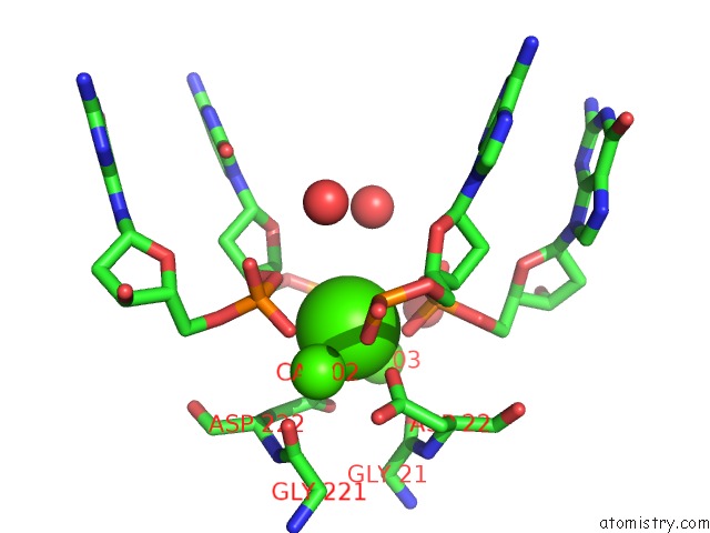
Mono view
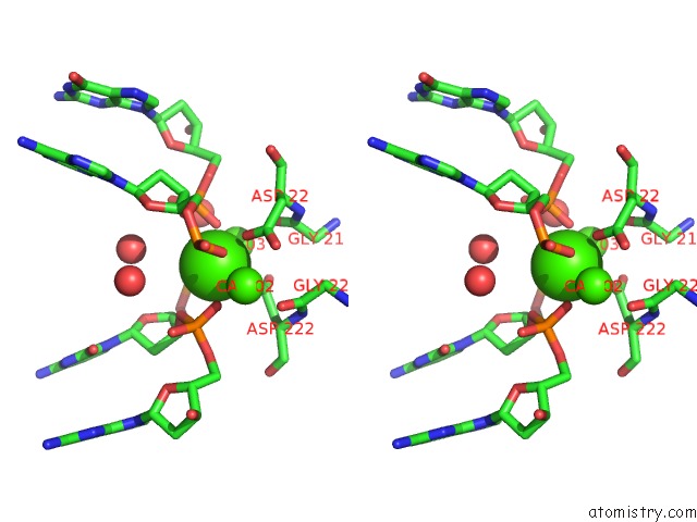
Stereo pair view
Mono view
Stereo pair view
A full contact list of Calcium with other atoms in the Ca binding
site number 1 of Crystal Structure of the Homing Endonuclease I-Msoi Bound to Its Dna Substrate within 5.0Å range:
|
Calcium binding site 2 out of 3 in 1m5x
Go back to
Calcium binding site 2 out
of 3 in the Crystal Structure of the Homing Endonuclease I-Msoi Bound to Its Dna Substrate
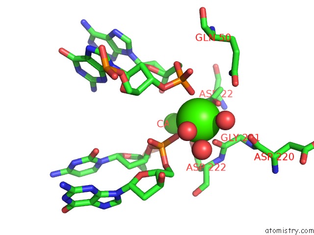
Mono view
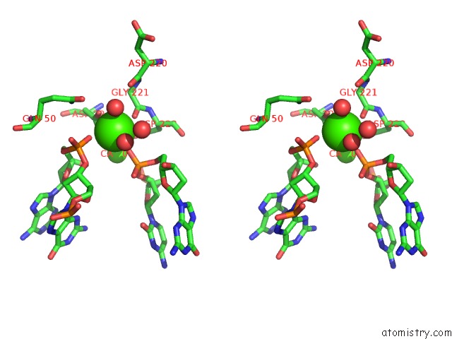
Stereo pair view
Mono view
Stereo pair view
A full contact list of Calcium with other atoms in the Ca binding
site number 2 of Crystal Structure of the Homing Endonuclease I-Msoi Bound to Its Dna Substrate within 5.0Å range:
|
Calcium binding site 3 out of 3 in 1m5x
Go back to
Calcium binding site 3 out
of 3 in the Crystal Structure of the Homing Endonuclease I-Msoi Bound to Its Dna Substrate

Mono view

Stereo pair view
Mono view
Stereo pair view
A full contact list of Calcium with other atoms in the Ca binding
site number 3 of Crystal Structure of the Homing Endonuclease I-Msoi Bound to Its Dna Substrate within 5.0Å range:
|
Reference:
B.Chevalier,
M.Turmel,
C.Lemieux,
R.J.Monnat,
B.L.Stoddard.
Flexible Dna Target Site Recognition By Divergent Homing Endonuclease Isoschizomers I-Crei and I-Msoi J.Mol.Biol. V. 329 253 2003.
ISSN: ISSN 0022-2836
PubMed: 12758074
DOI: 10.1016/S0022-2836(03)00447-9
Page generated: Thu Jul 11 12:16:22 2024
ISSN: ISSN 0022-2836
PubMed: 12758074
DOI: 10.1016/S0022-2836(03)00447-9
Last articles
Zn in 9JYWZn in 9IR4
Zn in 9IR3
Zn in 9GMX
Zn in 9GMW
Zn in 9JEJ
Zn in 9ERF
Zn in 9ERE
Zn in 9EGV
Zn in 9EGW