Calcium »
PDB 1lu1-1m8v »
1m8t »
Calcium in PDB 1m8t: Structure of An Acidic Phospholipase A2 From the Venom of Ophiophagus Hannah at 2.1 Resolution From A Hemihedrally Twinned Crystal Form
Enzymatic activity of Structure of An Acidic Phospholipase A2 From the Venom of Ophiophagus Hannah at 2.1 Resolution From A Hemihedrally Twinned Crystal Form
All present enzymatic activity of Structure of An Acidic Phospholipase A2 From the Venom of Ophiophagus Hannah at 2.1 Resolution From A Hemihedrally Twinned Crystal Form:
3.1.1.4;
3.1.1.4;
Protein crystallography data
The structure of Structure of An Acidic Phospholipase A2 From the Venom of Ophiophagus Hannah at 2.1 Resolution From A Hemihedrally Twinned Crystal Form, PDB code: 1m8t
was solved by
S.Xu,
L.Gu,
Q.Wang,
Y.Shu,
Z.Lin,
with X-Ray Crystallography technique. A brief refinement statistics is given in the table below:
| Resolution Low / High (Å) | 16.99 / 2.10 |
| Space group | P 63 |
| Cell size a, b, c (Å), α, β, γ (°) | 98.060, 98.060, 132.390, 90.00, 90.00, 120.00 |
| R / Rfree (%) | 19.2 / 21.3 |
Calcium Binding Sites:
The binding sites of Calcium atom in the Structure of An Acidic Phospholipase A2 From the Venom of Ophiophagus Hannah at 2.1 Resolution From A Hemihedrally Twinned Crystal Form
(pdb code 1m8t). This binding sites where shown within
5.0 Angstroms radius around Calcium atom.
In total 6 binding sites of Calcium where determined in the Structure of An Acidic Phospholipase A2 From the Venom of Ophiophagus Hannah at 2.1 Resolution From A Hemihedrally Twinned Crystal Form, PDB code: 1m8t:
Jump to Calcium binding site number: 1; 2; 3; 4; 5; 6;
In total 6 binding sites of Calcium where determined in the Structure of An Acidic Phospholipase A2 From the Venom of Ophiophagus Hannah at 2.1 Resolution From A Hemihedrally Twinned Crystal Form, PDB code: 1m8t:
Jump to Calcium binding site number: 1; 2; 3; 4; 5; 6;
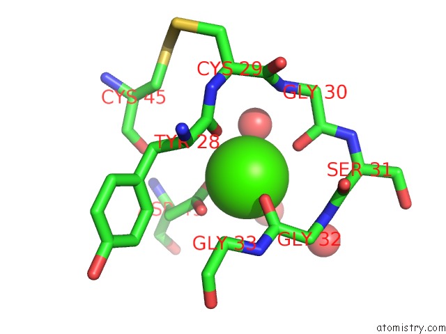
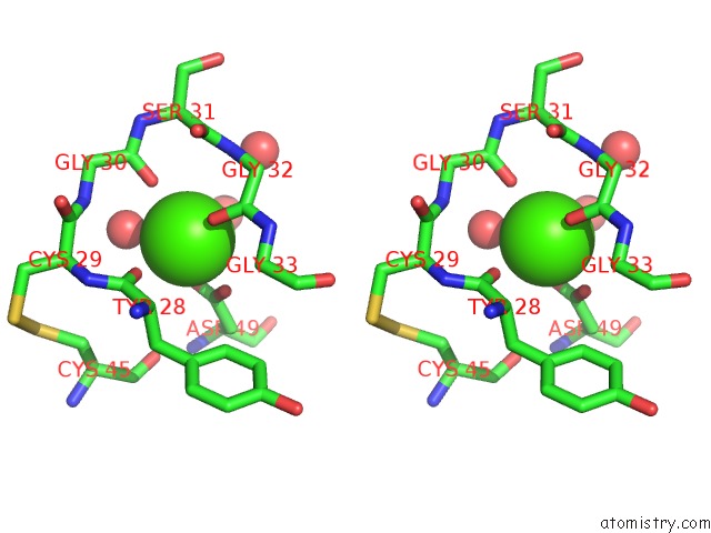
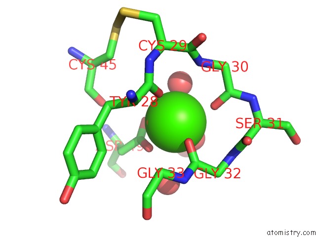
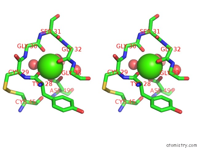
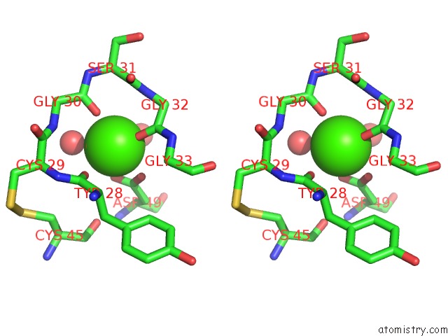
Calcium binding site 1 out of 6 in 1m8t
Go back to
Calcium binding site 1 out
of 6 in the Structure of An Acidic Phospholipase A2 From the Venom of Ophiophagus Hannah at 2.1 Resolution From A Hemihedrally Twinned Crystal Form
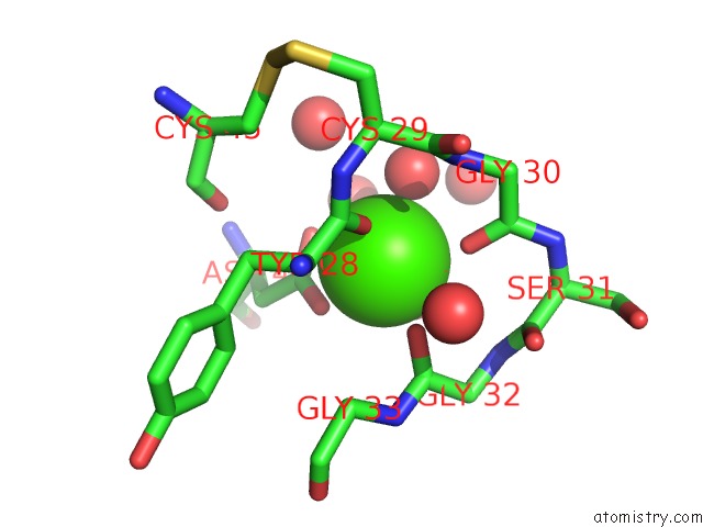
Mono view
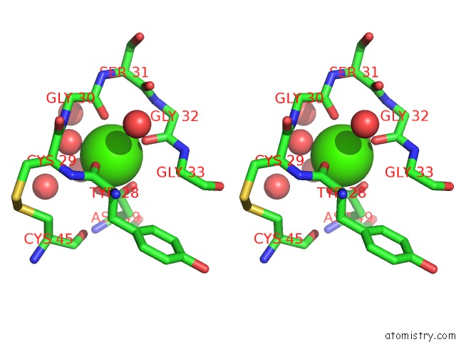
Stereo pair view
Mono view
Stereo pair view
A full contact list of Calcium with other atoms in the Ca binding
site number 1 of Structure of An Acidic Phospholipase A2 From the Venom of Ophiophagus Hannah at 2.1 Resolution From A Hemihedrally Twinned Crystal Form within 5.0Å range:
|
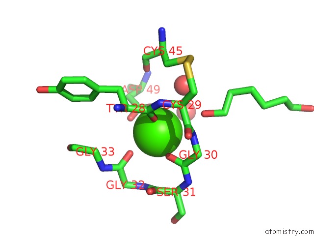
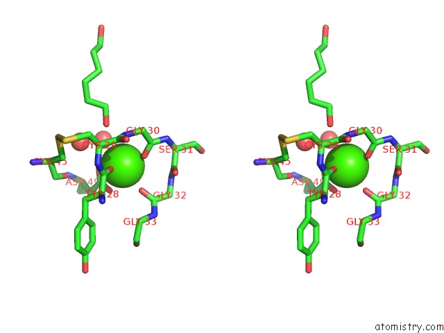
Calcium binding site 2 out of 6 in 1m8t
Go back to
Calcium binding site 2 out
of 6 in the Structure of An Acidic Phospholipase A2 From the Venom of Ophiophagus Hannah at 2.1 Resolution From A Hemihedrally Twinned Crystal Form
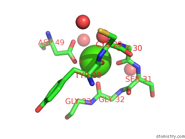
Mono view
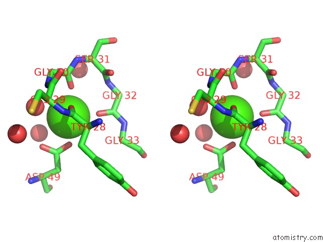
Stereo pair view
Mono view
Stereo pair view
A full contact list of Calcium with other atoms in the Ca binding
site number 2 of Structure of An Acidic Phospholipase A2 From the Venom of Ophiophagus Hannah at 2.1 Resolution From A Hemihedrally Twinned Crystal Form within 5.0Å range:
|
Calcium binding site 3 out of 6 in 1m8t
Go back to
Calcium binding site 3 out
of 6 in the Structure of An Acidic Phospholipase A2 From the Venom of Ophiophagus Hannah at 2.1 Resolution From A Hemihedrally Twinned Crystal Form
Mono view
Stereo pair view
Mono view
Stereo pair view
A full contact list of Calcium with other atoms in the Ca binding
site number 3 of Structure of An Acidic Phospholipase A2 From the Venom of Ophiophagus Hannah at 2.1 Resolution From A Hemihedrally Twinned Crystal Form within 5.0Å range:
|
Calcium binding site 4 out of 6 in 1m8t
Go back to
Calcium binding site 4 out
of 6 in the Structure of An Acidic Phospholipase A2 From the Venom of Ophiophagus Hannah at 2.1 Resolution From A Hemihedrally Twinned Crystal Form
Mono view
Stereo pair view
Mono view
Stereo pair view
A full contact list of Calcium with other atoms in the Ca binding
site number 4 of Structure of An Acidic Phospholipase A2 From the Venom of Ophiophagus Hannah at 2.1 Resolution From A Hemihedrally Twinned Crystal Form within 5.0Å range:
|
Calcium binding site 5 out of 6 in 1m8t
Go back to
Calcium binding site 5 out
of 6 in the Structure of An Acidic Phospholipase A2 From the Venom of Ophiophagus Hannah at 2.1 Resolution From A Hemihedrally Twinned Crystal Form
Mono view
Stereo pair view
Mono view
Stereo pair view
A full contact list of Calcium with other atoms in the Ca binding
site number 5 of Structure of An Acidic Phospholipase A2 From the Venom of Ophiophagus Hannah at 2.1 Resolution From A Hemihedrally Twinned Crystal Form within 5.0Å range:
|
Calcium binding site 6 out of 6 in 1m8t
Go back to
Calcium binding site 6 out
of 6 in the Structure of An Acidic Phospholipase A2 From the Venom of Ophiophagus Hannah at 2.1 Resolution From A Hemihedrally Twinned Crystal Form

Mono view
Stereo pair view
Mono view
Stereo pair view
A full contact list of Calcium with other atoms in the Ca binding
site number 6 of Structure of An Acidic Phospholipase A2 From the Venom of Ophiophagus Hannah at 2.1 Resolution From A Hemihedrally Twinned Crystal Form within 5.0Å range:
|
Reference:
S.Xu,
L.Gu,
Q.Wang,
Y.Shu,
S.Song,
Z.Lin.
Structure of A King Cobra Phospholipase A2 Determined From A Hemihedrally Twinned Crystal. Acta Crystallogr.,Sect.D V. 59 1574 2003.
ISSN: ISSN 0907-4449
PubMed: 12925787
DOI: 10.1107/S0907444903014598
Page generated: Thu Jul 11 12:20:20 2024
ISSN: ISSN 0907-4449
PubMed: 12925787
DOI: 10.1107/S0907444903014598
Last articles
Zn in 9JYWZn in 9IR4
Zn in 9IR3
Zn in 9GMX
Zn in 9GMW
Zn in 9JEJ
Zn in 9ERF
Zn in 9ERE
Zn in 9EGV
Zn in 9EGW