Calcium »
PDB 1lu1-1m8v »
1m8v »
Calcium in PDB 1m8v: Structure of Pyrococcus Abyssii Sm Protein in Complex with A Uridine Heptamer
Protein crystallography data
The structure of Structure of Pyrococcus Abyssii Sm Protein in Complex with A Uridine Heptamer, PDB code: 1m8v
was solved by
S.Thore,
C.Mayer,
C.Sauter,
S.Weeks,
D.Suck,
with X-Ray Crystallography technique. A brief refinement statistics is given in the table below:
| Resolution Low / High (Å) | 30.00 / 2.60 |
| Space group | P 1 |
| Cell size a, b, c (Å), α, β, γ (°) | 68.000, 68.000, 84.800, 105.00, 108.80, 100.00 |
| R / Rfree (%) | 21.2 / 28.2 |
Calcium Binding Sites:
The binding sites of Calcium atom in the Structure of Pyrococcus Abyssii Sm Protein in Complex with A Uridine Heptamer
(pdb code 1m8v). This binding sites where shown within
5.0 Angstroms radius around Calcium atom.
In total 7 binding sites of Calcium where determined in the Structure of Pyrococcus Abyssii Sm Protein in Complex with A Uridine Heptamer, PDB code: 1m8v:
Jump to Calcium binding site number: 1; 2; 3; 4; 5; 6; 7;
In total 7 binding sites of Calcium where determined in the Structure of Pyrococcus Abyssii Sm Protein in Complex with A Uridine Heptamer, PDB code: 1m8v:
Jump to Calcium binding site number: 1; 2; 3; 4; 5; 6; 7;
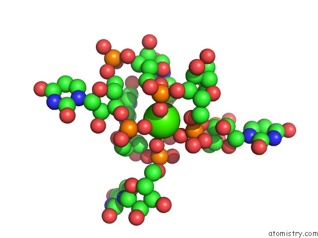
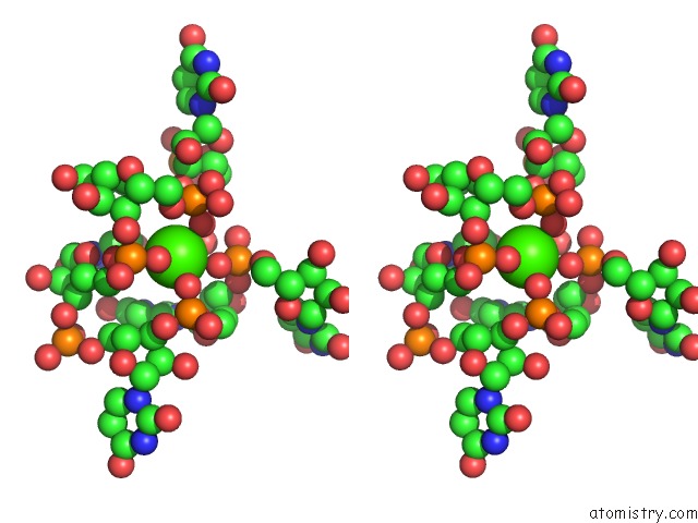
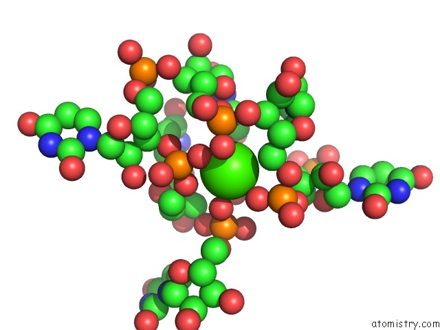
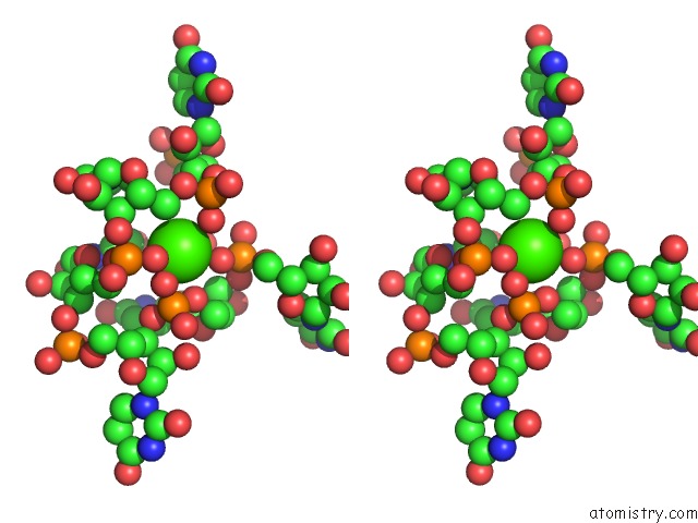
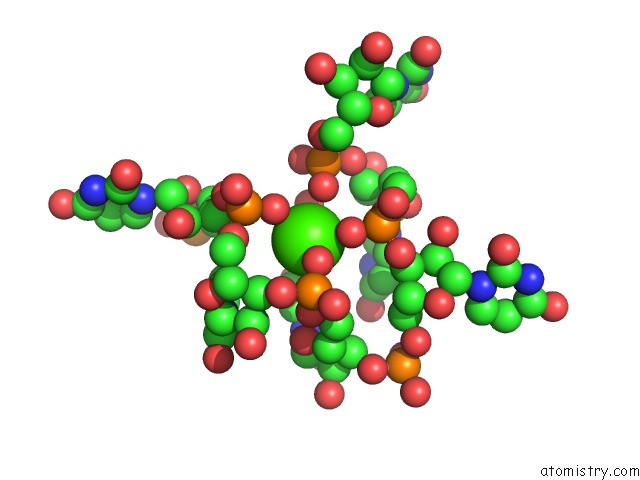
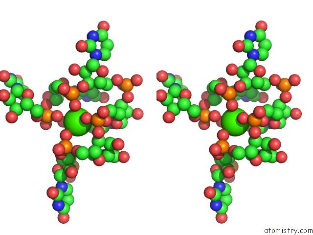
Calcium binding site 1 out of 7 in 1m8v
Go back to
Calcium binding site 1 out
of 7 in the Structure of Pyrococcus Abyssii Sm Protein in Complex with A Uridine Heptamer
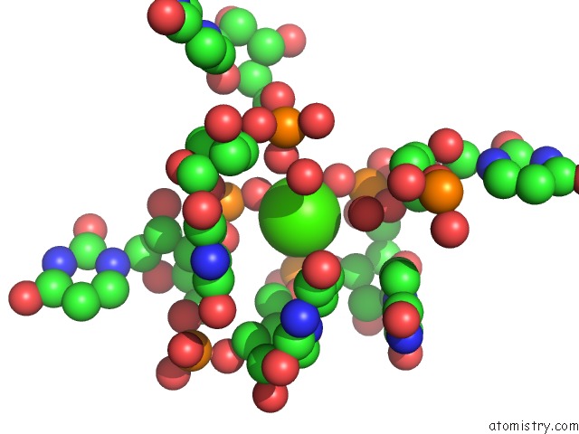
Mono view
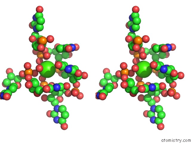
Stereo pair view
Mono view
Stereo pair view
A full contact list of Calcium with other atoms in the Ca binding
site number 1 of Structure of Pyrococcus Abyssii Sm Protein in Complex with A Uridine Heptamer within 5.0Å range:
|
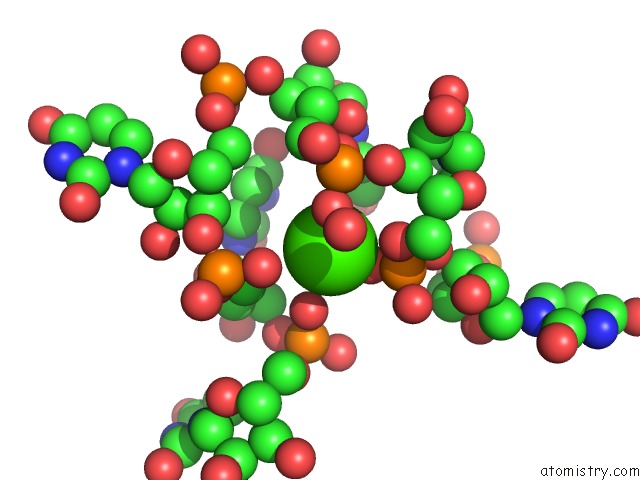
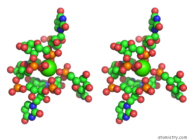
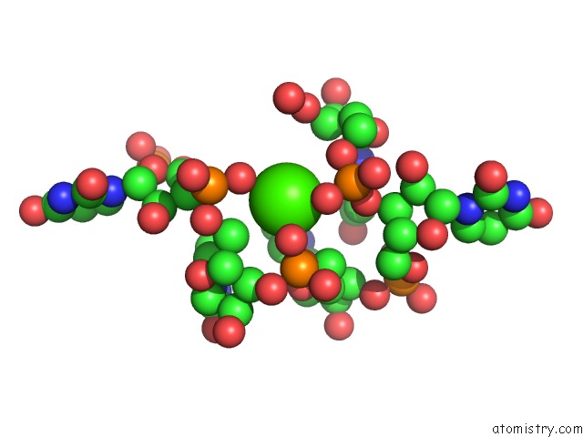
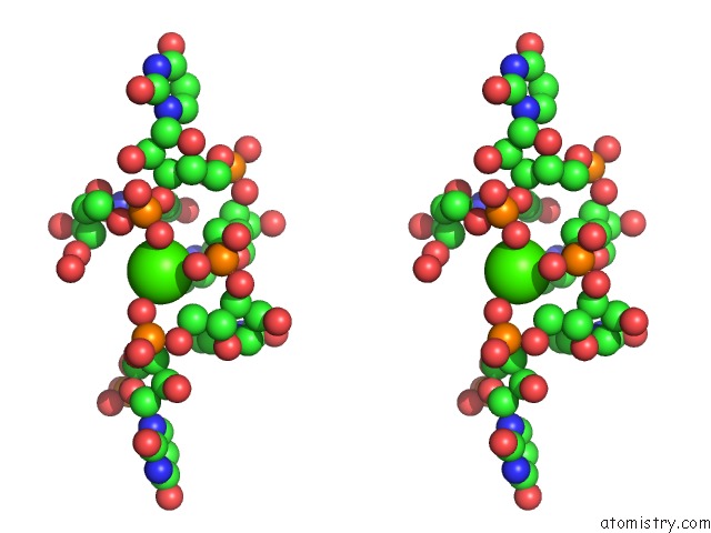
Calcium binding site 2 out of 7 in 1m8v
Go back to
Calcium binding site 2 out
of 7 in the Structure of Pyrococcus Abyssii Sm Protein in Complex with A Uridine Heptamer
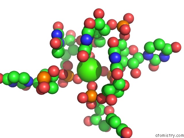
Mono view
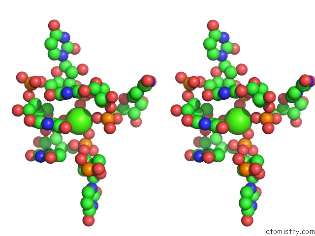
Stereo pair view
Mono view
Stereo pair view
A full contact list of Calcium with other atoms in the Ca binding
site number 2 of Structure of Pyrococcus Abyssii Sm Protein in Complex with A Uridine Heptamer within 5.0Å range:
|
Calcium binding site 3 out of 7 in 1m8v
Go back to
Calcium binding site 3 out
of 7 in the Structure of Pyrococcus Abyssii Sm Protein in Complex with A Uridine Heptamer
Mono view
Stereo pair view
Mono view
Stereo pair view
A full contact list of Calcium with other atoms in the Ca binding
site number 3 of Structure of Pyrococcus Abyssii Sm Protein in Complex with A Uridine Heptamer within 5.0Å range:
|
Calcium binding site 4 out of 7 in 1m8v
Go back to
Calcium binding site 4 out
of 7 in the Structure of Pyrococcus Abyssii Sm Protein in Complex with A Uridine Heptamer
Mono view
Stereo pair view
Mono view
Stereo pair view
A full contact list of Calcium with other atoms in the Ca binding
site number 4 of Structure of Pyrococcus Abyssii Sm Protein in Complex with A Uridine Heptamer within 5.0Å range:
|
Calcium binding site 5 out of 7 in 1m8v
Go back to
Calcium binding site 5 out
of 7 in the Structure of Pyrococcus Abyssii Sm Protein in Complex with A Uridine Heptamer
Mono view
Stereo pair view
Mono view
Stereo pair view
A full contact list of Calcium with other atoms in the Ca binding
site number 5 of Structure of Pyrococcus Abyssii Sm Protein in Complex with A Uridine Heptamer within 5.0Å range:
|
Calcium binding site 6 out of 7 in 1m8v
Go back to
Calcium binding site 6 out
of 7 in the Structure of Pyrococcus Abyssii Sm Protein in Complex with A Uridine Heptamer
Mono view
Stereo pair view
Mono view
Stereo pair view
A full contact list of Calcium with other atoms in the Ca binding
site number 6 of Structure of Pyrococcus Abyssii Sm Protein in Complex with A Uridine Heptamer within 5.0Å range:
|
Calcium binding site 7 out of 7 in 1m8v
Go back to
Calcium binding site 7 out
of 7 in the Structure of Pyrococcus Abyssii Sm Protein in Complex with A Uridine Heptamer
Mono view
Stereo pair view
Mono view
Stereo pair view
A full contact list of Calcium with other atoms in the Ca binding
site number 7 of Structure of Pyrococcus Abyssii Sm Protein in Complex with A Uridine Heptamer within 5.0Å range:
|
Reference:
S.Thore,
C.Mayer,
C.Sauter,
S.Weeks,
D.Suck.
Crystal Structure of Pyrococcus Abyssii Sm Core and Its Complex with Rna: Common Features of Rna-Binding in Archaea and Eukarya J.Biol.Chem. V. 278 1239 2003.
ISSN: ISSN 0021-9258
PubMed: 12409299
DOI: 10.1074/JBC.M207685200
Page generated: Thu Jul 11 12:23:34 2024
ISSN: ISSN 0021-9258
PubMed: 12409299
DOI: 10.1074/JBC.M207685200
Last articles
Zn in 9JYWZn in 9IR4
Zn in 9IR3
Zn in 9GMX
Zn in 9GMW
Zn in 9JEJ
Zn in 9ERF
Zn in 9ERE
Zn in 9EGV
Zn in 9EGW